

484
S. Biswas et al.
high-definition IR imaging in the laboratory using minimally-modified commercial
instruments. this is obviously more accessible and cost effective compared to a
synchrotron. this dual capability in the community, of rapid imaging using a synchrotron and slower laboratory based systems is likely to prove useful for many
practitioners and, doubtless, numerous new studies will appear using the highdefinition approach. We anticipate that this optimal configuration will become a
standard approach for imaging in the mid-IR.
16.3.2 Sampling Modes for Acquiring IR Imaging Data
While we have considered the design of the microscope and optimal configurations,
one of the most critical aspects of conducting IR imaging experiments is the manner in which light interacts with the sample. there are three most commonly used
modes for acquiring IR data. these are in transmission, transmission-reflection and
attenuated total reflectance (AtR) mode.
16.3.2.1 Transmission Mode
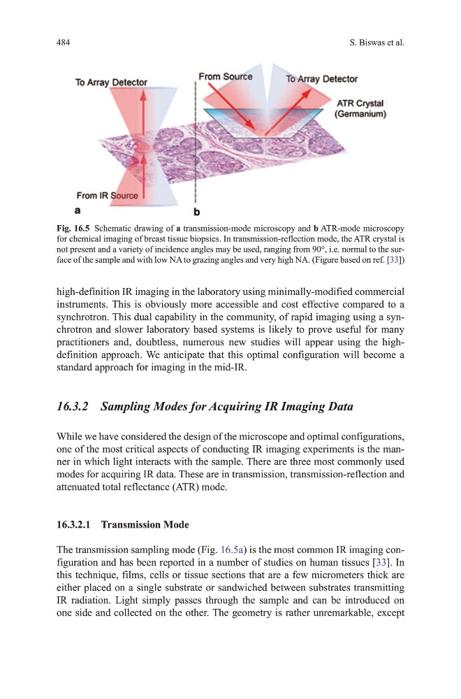
the transmission sampling mode (Fig. 16.5a) is the most common IR imaging configuration and has been reported in a number of studies on human tissues [33]. In
this technique, films, cells or tissue sections that are a few micrometers thick are
either placed on a single substrate or sandwiched between substrates transmitting
IR radiation. Light simply passes through the sample and can be introduced on
one side and collected on the other. the geometry is rather unremarkable, except
Fig. 16.5 Schematic drawing of a transmission-mode microscopy and b AtR-mode microscopy
for chemical imaging of breast tissue biopsies. In transmission-reflection mode, the AtR crystal is
not present and a variety of incidence angles may be used, ranging from 90°, i.e. normal to the surface of the sample and with low NA to grazing angles and very high NA. (Figure based on ref. [33])
S. Biswas et al.
high-definition IR imaging in the laboratory using minimally-modified commercial
instruments. this is obviously more accessible and cost effective compared to a
synchrotron. this dual capability in the community, of rapid imaging using a synchrotron and slower laboratory based systems is likely to prove useful for many
practitioners and, doubtless, numerous new studies will appear using the highdefinition approach. We anticipate that this optimal configuration will become a
standard approach for imaging in the mid-IR.
16.3.2 Sampling Modes for Acquiring IR Imaging Data
While we have considered the design of the microscope and optimal configurations,
one of the most critical aspects of conducting IR imaging experiments is the manner in which light interacts with the sample. there are three most commonly used
modes for acquiring IR data. these are in transmission, transmission-reflection and
attenuated total reflectance (AtR) mode.
16.3.2.1 Transmission Mode
the transmission sampling mode (Fig. 16.5a) is the most common IR imaging configuration and has been reported in a number of studies on human tissues [33]. In
this technique, films, cells or tissue sections that are a few micrometers thick are
either placed on a single substrate or sandwiched between substrates transmitting
IR radiation. Light simply passes through the sample and can be introduced on
one side and collected on the other. the geometry is rather unremarkable, except
Fig. 16.5 Schematic drawing of a transmission-mode microscopy and b AtR-mode microscopy
for chemical imaging of breast tissue biopsies. In transmission-reflection mode, the AtR crystal is
not present and a variety of incidence angles may be used, ranging from 90°, i.e. normal to the surface of the sample and with low NA to grazing angles and very high NA. (Figure based on ref. [33])