

483
16 Fourier Transform Infrared (Ft-Ir) Spectroscopic Imaging …
Another, more rigorous approach to understanding the quality of image formation was proposed using a Fourier optics approach [27]. A first principles understanding of the achievable quality as a function of instrument parameters was proposed, primarily to affect improved instrument design. A model for light propagation through an IR spectroscopic imaging system was first developed using scalar
wave theory. the model analytically describes the propagation of light along the
entire beam path between the source and the detector, while explicitly modeling
each component in the beam path. the effect of all optical elements and the sample
in the microscope was understood in terms of the accessible spatial frequencies in
the Fourier optics framework. Subsequently, simulations were conducted to explicitly obtain the image that would be recorded under certain conditions. the modeled
and measured images for both low NA and high NA systems, at specific detector
pixel spacing agreed remarkably. From this validated model, the optimal pixel size
at the sample plane is calculated and shown to be much smaller than that in current mid-infrared microscopy systems but larger than that obtained for the IRENI
system. A plot for the optimal pixel size as a function of optics and of wavelength
was provided such that any optimization point could be chosen rigorously. A commercial imaging system was then modified and experimental data were presented
to validate the developed model. Notably, this demonstration was accomplished on
a commercial benchtop IR imaging instrument. Without the use of a synchrotron,
acquired data were of high spatial quality but, as expected, of poorer SNR. Signal
processing approaches were implemented to improve the spectral SNR. At present, a number of studies have produced data demonstrating the ability to perform
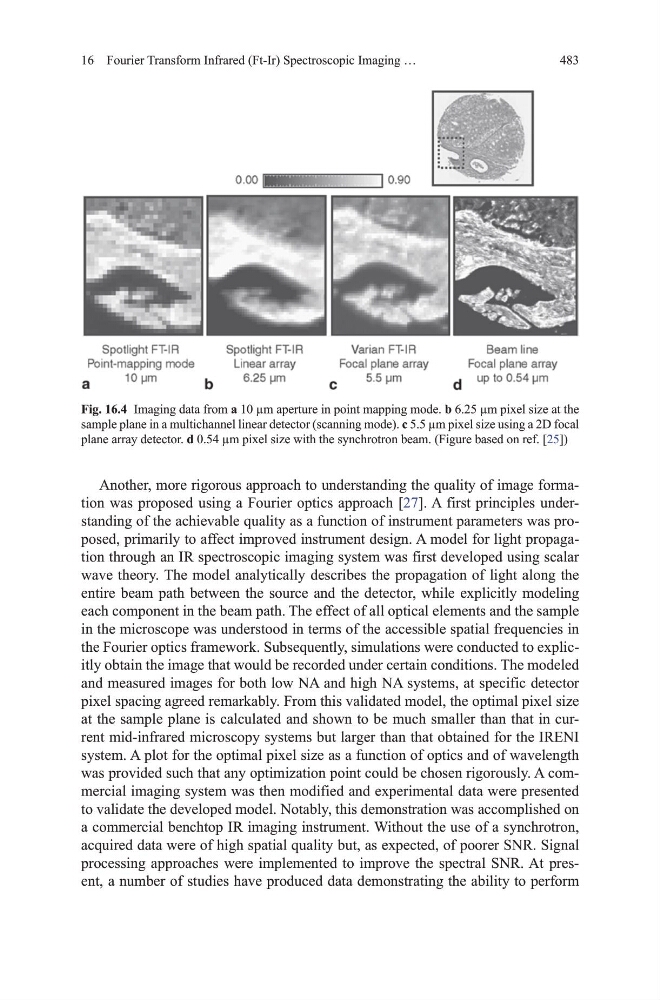
Fig. 16.4 Imaging data from a 10 µm aperture in point mapping mode. b 6.25 µm pixel size at the
sample plane in a multichannel linear detector (scanning mode). c 5.5 µm pixel size using a 2d focal
plane array detector. d 0.54 µm pixel size with the synchrotron beam. (Figure based on ref. [25])
16 Fourier Transform Infrared (Ft-Ir) Spectroscopic Imaging …
Another, more rigorous approach to understanding the quality of image formation was proposed using a Fourier optics approach [27]. A first principles understanding of the achievable quality as a function of instrument parameters was proposed, primarily to affect improved instrument design. A model for light propagation through an IR spectroscopic imaging system was first developed using scalar
wave theory. the model analytically describes the propagation of light along the
entire beam path between the source and the detector, while explicitly modeling
each component in the beam path. the effect of all optical elements and the sample
in the microscope was understood in terms of the accessible spatial frequencies in
the Fourier optics framework. Subsequently, simulations were conducted to explicitly obtain the image that would be recorded under certain conditions. the modeled
and measured images for both low NA and high NA systems, at specific detector
pixel spacing agreed remarkably. From this validated model, the optimal pixel size
at the sample plane is calculated and shown to be much smaller than that in current mid-infrared microscopy systems but larger than that obtained for the IRENI
system. A plot for the optimal pixel size as a function of optics and of wavelength
was provided such that any optimization point could be chosen rigorously. A commercial imaging system was then modified and experimental data were presented
to validate the developed model. Notably, this demonstration was accomplished on
a commercial benchtop IR imaging instrument. Without the use of a synchrotron,
acquired data were of high spatial quality but, as expected, of poorer SNR. Signal
processing approaches were implemented to improve the spectral SNR. At present, a number of studies have produced data demonstrating the ability to perform
Fig. 16.4 Imaging data from a 10 µm aperture in point mapping mode. b 6.25 µm pixel size at the
sample plane in a multichannel linear detector (scanning mode). c 5.5 µm pixel size using a 2d focal
plane array detector. d 0.54 µm pixel size with the synchrotron beam. (Figure based on ref. [25])