

3 Surface-enhanced Raman Scattering ...
37
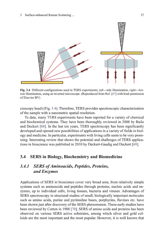
croscopy head) (Fig. 3.4). therefore, tERS provides spectroscopic characterization
of the sample with a nanometric spatial resolution.
to date, many tERS experiments have been reported for a variety of chemical
and biochemical systems. they have been thoroughly reviewed in 2008 by Bailo
and deckert [68]. In the last ten years, tERS spectroscopy has been significantly
developed and opened new possibilities of applications in a variety of fields in biology and medicine. In particular, experiments with living cells seem to be very promising. Interesting review that shows the potential and challenges of tERS applications in bioscience was published in 2010 by deckert-gaudig and deckert [69].
3.4 SERS in Biology, Biochemistry and Biomedicine
3.4.1 SERS of Aminoacids, Peptides, Proteins,
and Enzymes
Applications of SERS in bioscience cover very broad area, from relatively simple
systems such as aminoacids and peptides through proteins, nucleic acids and enzymes, up to individual cells, living tissues, bacteria and viruses. Advantages of
SERS spectroscopy in structural studies of small, biologically important molecules
such as amino acids, purine and pyrimidine bases, porphyrins, flavines etc. have
been shown just after discovery of the SERS phenomenon. these early studies have
been reviewed by Cotton in 1988 [70]. SERS of amino acids and proteins has been
observed on various SERS active substrates, among which silver and gold colloids are the most important and the most popular. however, it is well known that
Fig. 3.4 different configurations used in tERS experiments; left—side illumination, right—bottom illumination, using an inverted microscope. (Reproduced from Ref. [67] with kind permission
of Elsevier Bv)
37
croscopy head) (Fig. 3.4). therefore, tERS provides spectroscopic characterization
of the sample with a nanometric spatial resolution.
to date, many tERS experiments have been reported for a variety of chemical
and biochemical systems. they have been thoroughly reviewed in 2008 by Bailo
and deckert [68]. In the last ten years, tERS spectroscopy has been significantly
developed and opened new possibilities of applications in a variety of fields in biology and medicine. In particular, experiments with living cells seem to be very promising. Interesting review that shows the potential and challenges of tERS applications in bioscience was published in 2010 by deckert-gaudig and deckert [69].
3.4 SERS in Biology, Biochemistry and Biomedicine
3.4.1 SERS of Aminoacids, Peptides, Proteins,
and Enzymes
Applications of SERS in bioscience cover very broad area, from relatively simple
systems such as aminoacids and peptides through proteins, nucleic acids and enzymes, up to individual cells, living tissues, bacteria and viruses. Advantages of
SERS spectroscopy in structural studies of small, biologically important molecules
such as amino acids, purine and pyrimidine bases, porphyrins, flavines etc. have
been shown just after discovery of the SERS phenomenon. these early studies have
been reviewed by Cotton in 1988 [70]. SERS of amino acids and proteins has been
observed on various SERS active substrates, among which silver and gold colloids are the most important and the most popular. however, it is well known that
Fig. 3.4 different configurations used in tERS experiments; left—side illumination, right—bottom illumination, using an inverted microscope. (Reproduced from Ref. [67] with kind permission
of Elsevier Bv)