

J. Bukowska and P. Piotrowski
36
from the environment, preserving high enhancement factor of the shell-isolated
nanoparticles (ShINERS—shell-isolated nanoparticle enhanced Raman spectroscopy). Protective layers of silica [54], glass [55] or polyethylene glycol [56] are also
applied for metallic nanoparticles labeled with various compounds called Raman
reporters. these encapsulated nanoprobes find many applications as SERS probes
incorporated in a variety of biological materials (see Sect. 4.2). Larger enhancement
of the Raman scattering than that produced by a single nanoparticle (NP) is created
at the junction between two nanoparticles in a dimer, in which nanoparticles are
separated by nanometer-scale gaps [57–60]. Such NP dimers are sometimes called
hot spots. Reproducible fabrication of these nanostructures is however challenging,
because the SERS enhancement factor critically depends on interparticle spacing
[61].
this short review does not cover all developed SERS supports. usually, SERS
substrates are prepared in a given laboratory, but effectively enhancing gold solid
support known as Klarite, or colloid suspensions are now also commercially available.
Another approach to enhance Raman scattering is offered by tip-enhanced Raman spectroscopy (tERS), reported for the first time in 2000 [62] and significantly
developed in the last ten years [63–67]. In tERS, very strong electromagnetic field
is located at the nanometer-scale metallic tip (Ag or Au). In an ideal case, it ends
with a single metal nanoparticle. Its radius is much smaller than the diffraction limit
so it allows measuring the spectrum from a much more confined spot. the Raman
signal is strongly enhanced when tip is brought close to the sample irradiated by the
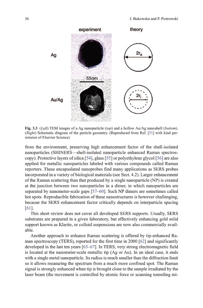
laser beam (the movement is controlled by atomic force or scanning tunneling miFig. 3.3 ( Left) tEm images of a Ag nanoparticle ( top) and a hollow Au/Ag nanoshell ( bottom).
( Right) Schematic diagram of the particle geometry. (Reproduced from Ref. [51] with kind permission of Elsevier Science)
36
from the environment, preserving high enhancement factor of the shell-isolated
nanoparticles (ShINERS—shell-isolated nanoparticle enhanced Raman spectroscopy). Protective layers of silica [54], glass [55] or polyethylene glycol [56] are also
applied for metallic nanoparticles labeled with various compounds called Raman
reporters. these encapsulated nanoprobes find many applications as SERS probes
incorporated in a variety of biological materials (see Sect. 4.2). Larger enhancement
of the Raman scattering than that produced by a single nanoparticle (NP) is created
at the junction between two nanoparticles in a dimer, in which nanoparticles are
separated by nanometer-scale gaps [57–60]. Such NP dimers are sometimes called
hot spots. Reproducible fabrication of these nanostructures is however challenging,
because the SERS enhancement factor critically depends on interparticle spacing
[61].
this short review does not cover all developed SERS supports. usually, SERS
substrates are prepared in a given laboratory, but effectively enhancing gold solid
support known as Klarite, or colloid suspensions are now also commercially available.
Another approach to enhance Raman scattering is offered by tip-enhanced Raman spectroscopy (tERS), reported for the first time in 2000 [62] and significantly
developed in the last ten years [63–67]. In tERS, very strong electromagnetic field
is located at the nanometer-scale metallic tip (Ag or Au). In an ideal case, it ends
with a single metal nanoparticle. Its radius is much smaller than the diffraction limit
so it allows measuring the spectrum from a much more confined spot. the Raman
signal is strongly enhanced when tip is brought close to the sample irradiated by the
laser beam (the movement is controlled by atomic force or scanning tunneling miFig. 3.3 ( Left) tEm images of a Ag nanoparticle ( top) and a hollow Au/Ag nanoshell ( bottom).
( Right) Schematic diagram of the particle geometry. (Reproduced from Ref. [51] with kind permission of Elsevier Science)