

454
K. malek et al.
microspectroscopy [126]. FtIR mapping of a tissue section derived from autopsy
material showed a significant change in the amide I wavenumber in the region of
neuritic plaques identified earlier from a histological examination. the position of
this band shifts from 1652–1654 cm
−1
in the periphery of the plaque to 1632–
1634 cm
−1
in its center. this clearly indicates a substantially different conformation
of proteins in abnormal deposit in the Ad tissue, suggesting an elevated level of
β-sheet proteins. The position of the plaque protein found in the tissue differed from
the position of synthetic amyloid (1620–1628 cm
−1
), however, this fact can result
from packing of other proteins around the amyloid deposit that may influence the
structure of the aggregated β-amyloid [126]. the spectral features associated with
an elevated content of β-sheet proteins in ca. 30 plaques of the human Ad brain tissue has been confirmed by miller et al. [127]. In this case, the position of the amide
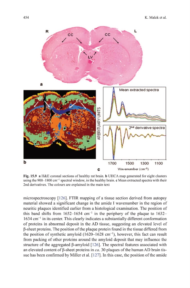
Fig. 15.9 a h&E coronal sections of healthy rat brain. b uhCA map generated for eight clusters
using the 900–1800 cm
−1
spectral window, in the healthy brain. c mean extracted spectra with their
2nd derivatives. the colours are explained in the main text
K. malek et al.
microspectroscopy [126]. FtIR mapping of a tissue section derived from autopsy
material showed a significant change in the amide I wavenumber in the region of
neuritic plaques identified earlier from a histological examination. the position of
this band shifts from 1652–1654 cm
−1
in the periphery of the plaque to 1632–
1634 cm
−1
in its center. this clearly indicates a substantially different conformation
of proteins in abnormal deposit in the Ad tissue, suggesting an elevated level of
β-sheet proteins. The position of the plaque protein found in the tissue differed from
the position of synthetic amyloid (1620–1628 cm
−1
), however, this fact can result
from packing of other proteins around the amyloid deposit that may influence the
structure of the aggregated β-amyloid [126]. the spectral features associated with
an elevated content of β-sheet proteins in ca. 30 plaques of the human Ad brain tissue has been confirmed by miller et al. [127]. In this case, the position of the amide
Fig. 15.9 a h&E coronal sections of healthy rat brain. b uhCA map generated for eight clusters
using the 900–1800 cm
−1
spectral window, in the healthy brain. c mean extracted spectra with their
2nd derivatives. the colours are explained in the main text