

J. Bukowska and P. Piotrowski
34
Soon after discovery of the SERS effect, metal (Ag and Au) colloids have been
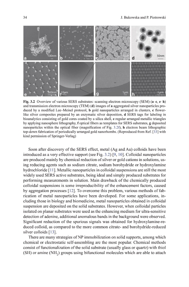
introduced as a very effective support (see Fig. 3.2) [9, 10]. Colloidal nanoparticles
are produced mainly by chemical reduction of silver or gold cations in solutions, using reducing agents such as sodium citrate, sodium borohydride or hydroxylamine
hydrochloride [11]. metallic nanoparticles in colloidal suspensions are still the most
widely used SERS active substrates, being ideal and simply produced substrates for
performing measurements in solution. main drawback of the chemically produced
colloidal suspensions is some irreproducibility of the enhancement factors, caused
by aggregation processes [12]. to overcome this problem, various methods of fabrication of metal nanoparticles have been developed. For some applications, including those in biology and biomedicine, metal nanoparticles obtained in colloidal
suspension are deposited on the solid substrates. however, when colloidal particles
isolated on planar substrates were used as the enhancing medium for ultra-sensitive
detection of adenine, additional anomalous bands in the background were observed.
Significant reduction of the spurious signals was obtained for hydroxylamine-reduced colloid, as compared to the more common citrate- and borohydride-reduced
silver colloids [13].
there are many strategies of NP immobilization on solid supports, among which
chemical or electrostatic self-assembling are the most popular. Chemical methods
consist of functionalization of the solid substrate (usually glass or quartz) with thiol
(Sh) or amine (Nh 2 ) groups using bifunctional molecules which are able to attach
Fig. 3.2 overview of various SERS substrates: scanning electron microscopy (SEm) (a–c, e–h)
and transmission electron microscopy (tEm) (d) images of a aggregated silver nanoparticles produced by a modified Lee–meisel protocol, b gold nanoparticles arranged in clusters, c flowerlike silver composites prepared by an enzymatic silver deposition, d SERS tags for labeling in
bioanalytics consisting of gold cores coated by a silica shell, e regular arranged metallic triangles
by applying nanosphere lithography, f optical fibers as templates for SERS substrates, g deposited
nanoparticles within the optical fiber (magnification of Fig. 3.2f), h electron beam lithographic
top-down fabrication of periodically arranged gold nanorhombs. (Reproduced from Ref. [33] with
kind permission of Springer-verlag)
34
Soon after discovery of the SERS effect, metal (Ag and Au) colloids have been
introduced as a very effective support (see Fig. 3.2) [9, 10]. Colloidal nanoparticles
are produced mainly by chemical reduction of silver or gold cations in solutions, using reducing agents such as sodium citrate, sodium borohydride or hydroxylamine
hydrochloride [11]. metallic nanoparticles in colloidal suspensions are still the most
widely used SERS active substrates, being ideal and simply produced substrates for
performing measurements in solution. main drawback of the chemically produced
colloidal suspensions is some irreproducibility of the enhancement factors, caused
by aggregation processes [12]. to overcome this problem, various methods of fabrication of metal nanoparticles have been developed. For some applications, including those in biology and biomedicine, metal nanoparticles obtained in colloidal
suspension are deposited on the solid substrates. however, when colloidal particles
isolated on planar substrates were used as the enhancing medium for ultra-sensitive
detection of adenine, additional anomalous bands in the background were observed.
Significant reduction of the spurious signals was obtained for hydroxylamine-reduced colloid, as compared to the more common citrate- and borohydride-reduced
silver colloids [13].
there are many strategies of NP immobilization on solid supports, among which
chemical or electrostatic self-assembling are the most popular. Chemical methods
consist of functionalization of the solid substrate (usually glass or quartz) with thiol
(Sh) or amine (Nh 2 ) groups using bifunctional molecules which are able to attach
Fig. 3.2 overview of various SERS substrates: scanning electron microscopy (SEm) (a–c, e–h)
and transmission electron microscopy (tEm) (d) images of a aggregated silver nanoparticles produced by a modified Lee–meisel protocol, b gold nanoparticles arranged in clusters, c flowerlike silver composites prepared by an enzymatic silver deposition, d SERS tags for labeling in
bioanalytics consisting of gold cores coated by a silica shell, e regular arranged metallic triangles
by applying nanosphere lithography, f optical fibers as templates for SERS substrates, g deposited
nanoparticles within the optical fiber (magnification of Fig. 3.2f), h electron beam lithographic
top-down fabrication of periodically arranged gold nanorhombs. (Reproduced from Ref. [33] with
kind permission of Springer-verlag)