

387
13 Vibrational Spectroscopy: Disease Diagnostics and Beyond
are greatly enhanced by the minimal need for data-preprocessing and prolonged
measurement periods. these conditions are facilitated by the use of collagen 3-d
matrices for cell growth and measurement.
13.6 NanoParticles in Cells [151]
the potential of vibrational, particularly Raman, spectroscopy as a realtime label
free subcellular analytical technique opens up a range of applications, as for example in the field of nanotoxicology and nanomedicine. one of the major concerns regarding the possible toxic effects of nanoparticles is the capacity of these
materials to penetrate cells and possibly translocate to other cells around the body.
Conventional in vitro cytotoxicity assays, such as Alamar Blue, Neutral Red, mtt,
etc, provide indications of impact on cell proliferation, viability, metabolic activity,
lysosomal and mitochondrial activity. however, they fail to provide a verification
of nanoparticle internalisation or elucidate intracellular trafficking mechanisms and
subcellular distribution within cells. Indeed, false positive results have been demonstrated due to the extracellular interaction of the nanoparticles with the cell culture
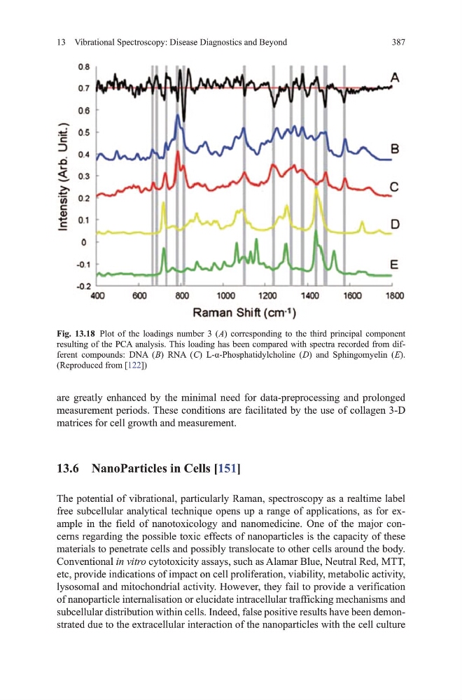
Fig. 13.18 Plot of the loadings number 3 ( A) corresponding to the third principal component
resulting of the PCA analysis. this loading has been compared with spectra recorded from different compounds: dNA ( B) RNA ( C) L-α-Phosphatidylcholine ( D) and Sphingomyelin ( E).
(Reproduced from [122])
13 Vibrational Spectroscopy: Disease Diagnostics and Beyond
are greatly enhanced by the minimal need for data-preprocessing and prolonged
measurement periods. these conditions are facilitated by the use of collagen 3-d
matrices for cell growth and measurement.
13.6 NanoParticles in Cells [151]
the potential of vibrational, particularly Raman, spectroscopy as a realtime label
free subcellular analytical technique opens up a range of applications, as for example in the field of nanotoxicology and nanomedicine. one of the major concerns regarding the possible toxic effects of nanoparticles is the capacity of these
materials to penetrate cells and possibly translocate to other cells around the body.
Conventional in vitro cytotoxicity assays, such as Alamar Blue, Neutral Red, mtt,
etc, provide indications of impact on cell proliferation, viability, metabolic activity,
lysosomal and mitochondrial activity. however, they fail to provide a verification
of nanoparticle internalisation or elucidate intracellular trafficking mechanisms and
subcellular distribution within cells. Indeed, false positive results have been demonstrated due to the extracellular interaction of the nanoparticles with the cell culture
Fig. 13.18 Plot of the loadings number 3 ( A) corresponding to the third principal component
resulting of the PCA analysis. this loading has been compared with spectra recorded from different compounds: dNA ( B) RNA ( C) L-α-Phosphatidylcholine ( D) and Sphingomyelin ( E).
(Reproduced from [122])