

368
H. J. Byrne et al.
cervical cancers as a result of functional inactivation of pRb by the hPv E7 protein
[77]. this overexpression highlights the potential of p16
INK4A
as a marker for CIN
and cervical cancer. hPv positivity and p16
INK4A
positivity have shown a correlation, even though p16
INK4A
expression was also seen in a limited number of hPv
negative biopsy samples [78]. Also, a correlation between p16
INK4A
expression and
cervical lesion grade and hR-hPv positivity has been documented [79]. p16
INK4A
has been proven to be the most reliable marker of cervical dysplasia and was found
to mark all grades of squamous and glandular lesions of the cervix. the use of
p16
INK4A
immunocytochemical analysis as a complement to conventional screening
programmes could potentially aid in the reduction of false positive and false negative results [80].
In the first part of this study, expression of p16
INK4A
was analysed in cervical cancer cell lines using immunocytochemical staining and both confocal fluorescence
microscopy and flow cytometry. [64] Fluorescence images of cells were collected
using a confocal fluorescence microscopy system. Representative images of cells
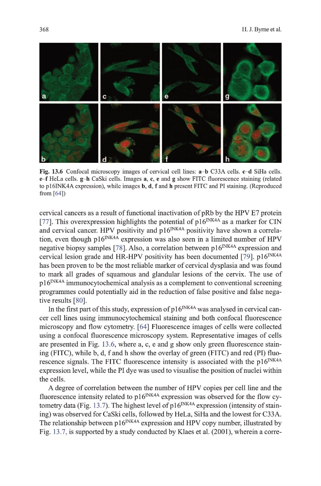
are presented in Fig. 13.6, where a, c, e and g show only green fluorescence staining (FItC), while b, d, f and h show the overlay of green (FItC) and red (PI) fluorescence signals. the FItC fluorescence intensity is associated with the p16
INK4A
expression level, while the PI dye was used to visualise the position of nuclei within
the cells.
A degree of correlation between the number of hPv copies per cell line and the
fluorescence intensity related to p16
INK4A
expression was observed for the flow cytometry data (Fig. 13.7). the highest level of p16
INK4A
expression (intensity of staining) was observed for CaSki cells, followed by heLa, Siha and the lowest for C33A.
the relationship between p16
INK4A
expression and hPv copy number, illustrated by
Fig. 13.7, is supported by a study conducted by Klaes et al. (2001), wherein a correFig. 13.6 Confocal microscopy images of cervical cell lines: a–b C33A cells. c–d Siha cells.
e–f heLa cells. g–h CaSki cells. Images a, c, e and g show FItC fluorescence staining (related
to p16INK4A expression), while images b, d, f and h present FItC and PI staining. (Reproduced
from [64])
H. J. Byrne et al.
cervical cancers as a result of functional inactivation of pRb by the hPv E7 protein
[77]. this overexpression highlights the potential of p16
INK4A
as a marker for CIN
and cervical cancer. hPv positivity and p16
INK4A
positivity have shown a correlation, even though p16
INK4A
expression was also seen in a limited number of hPv
negative biopsy samples [78]. Also, a correlation between p16
INK4A
expression and
cervical lesion grade and hR-hPv positivity has been documented [79]. p16
INK4A
has been proven to be the most reliable marker of cervical dysplasia and was found
to mark all grades of squamous and glandular lesions of the cervix. the use of
p16
INK4A
immunocytochemical analysis as a complement to conventional screening
programmes could potentially aid in the reduction of false positive and false negative results [80].
In the first part of this study, expression of p16
INK4A
was analysed in cervical cancer cell lines using immunocytochemical staining and both confocal fluorescence
microscopy and flow cytometry. [64] Fluorescence images of cells were collected
using a confocal fluorescence microscopy system. Representative images of cells
are presented in Fig. 13.6, where a, c, e and g show only green fluorescence staining (FItC), while b, d, f and h show the overlay of green (FItC) and red (PI) fluorescence signals. the FItC fluorescence intensity is associated with the p16
INK4A
expression level, while the PI dye was used to visualise the position of nuclei within
the cells.
A degree of correlation between the number of hPv copies per cell line and the
fluorescence intensity related to p16
INK4A
expression was observed for the flow cytometry data (Fig. 13.7). the highest level of p16
INK4A
expression (intensity of staining) was observed for CaSki cells, followed by heLa, Siha and the lowest for C33A.
the relationship between p16
INK4A
expression and hPv copy number, illustrated by
Fig. 13.7, is supported by a study conducted by Klaes et al. (2001), wherein a correFig. 13.6 Confocal microscopy images of cervical cell lines: a–b C33A cells. c–d Siha cells.
e–f heLa cells. g–h CaSki cells. Images a, c, e and g show FItC fluorescence staining (related
to p16INK4A expression), while images b, d, f and h present FItC and PI staining. (Reproduced
from [64])