

321
Additionally, the approximate concentration of carotenoids in single cells was estimated based on comparison of the Raman intensity of carotenoid signals in the
solution (known concentration) and cells (unknown concentration) [9]. Based on
the appearance of the marker bands at 1525 and 1157 cm
−1
in the Raman spectra,
the presence of carotenoids in the subcellular compartments was investigated. No
characteristic bands of carotenoids in the Raman spectra of nuclei, neither in B- nor
t-lymphocytes, nor NK-cells were found. on the contrary, the high concentration
of carotenoids was observed in some places in cytoplasm for t-lymphocytes and
NK-cells [9]. For both these types of cells, the particularly elevated concentration of
carotenoids was found in gall bodies (about 10
−3
mol dm
−3
) compared to the normal
physiological carotenoids concentration in the blood plasma of 10
−6
mol dm
−3
[53].
As expected, the reason of the high concentration of carotenoids predominantly in
the subcellular components containing high lipid concentration e.g. in gall bodies
is due to the lipophilic nature of these pigments [9]. distribution of carotenoids in
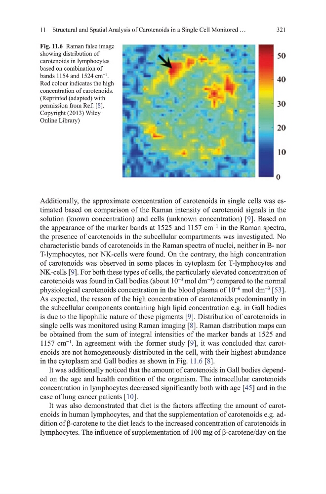
single cells was monitored using Raman imaging [8]. Raman distribution maps can
be obtained from the sum of integral intensities of the marker bands at 1525 and
1157 cm
−1
. In agreement with the former study [9], it was concluded that carotenoids are not homogeneously distributed in the cell, with their highest abundance
in the cytoplasm and gall bodies as shown in Fig. 11.6 [8].
It was additionally noticed that the amount of carotenoids in gall bodies depended on the age and health condition of the organism. the intracellular carotenoids
concentration in lymphocytes decreased significantly both with age [45] and in the
case of lung cancer patients [10].
It was also demonstrated that diet is the factors affecting the amount of carotenoids in human lymphocytes, and that the supplementation of carotenoids e.g. addition of β-carotene to the diet leads to the increased concentration of carotenoids in
lymphocytes. The influence of supplementation of 100 mg of β-carotene/day on the
Fig. 11.6 Raman false image
showing distribution of
carotenoids in lymphocytes
based on combination of
bands 1154 and 1524 cm
−1
.
Red colour indicates the high
concentration of carotenoids.
(Reprinted (adapted) with
permission from Ref. [8].
Copyright (2013) Wiley
online Library)
11 Structural and Spatial Analysis of Carotenoids in a Single Cell monitored …
Additionally, the approximate concentration of carotenoids in single cells was estimated based on comparison of the Raman intensity of carotenoid signals in the
solution (known concentration) and cells (unknown concentration) [9]. Based on
the appearance of the marker bands at 1525 and 1157 cm
−1
in the Raman spectra,
the presence of carotenoids in the subcellular compartments was investigated. No
characteristic bands of carotenoids in the Raman spectra of nuclei, neither in B- nor
t-lymphocytes, nor NK-cells were found. on the contrary, the high concentration
of carotenoids was observed in some places in cytoplasm for t-lymphocytes and
NK-cells [9]. For both these types of cells, the particularly elevated concentration of
carotenoids was found in gall bodies (about 10
−3
mol dm
−3
) compared to the normal
physiological carotenoids concentration in the blood plasma of 10
−6
mol dm
−3
[53].
As expected, the reason of the high concentration of carotenoids predominantly in
the subcellular components containing high lipid concentration e.g. in gall bodies
is due to the lipophilic nature of these pigments [9]. distribution of carotenoids in
single cells was monitored using Raman imaging [8]. Raman distribution maps can
be obtained from the sum of integral intensities of the marker bands at 1525 and
1157 cm
−1
. In agreement with the former study [9], it was concluded that carotenoids are not homogeneously distributed in the cell, with their highest abundance
in the cytoplasm and gall bodies as shown in Fig. 11.6 [8].
It was additionally noticed that the amount of carotenoids in gall bodies depended on the age and health condition of the organism. the intracellular carotenoids
concentration in lymphocytes decreased significantly both with age [45] and in the
case of lung cancer patients [10].
It was also demonstrated that diet is the factors affecting the amount of carotenoids in human lymphocytes, and that the supplementation of carotenoids e.g. addition of β-carotene to the diet leads to the increased concentration of carotenoids in
lymphocytes. The influence of supplementation of 100 mg of β-carotene/day on the
Fig. 11.6 Raman false image
showing distribution of
carotenoids in lymphocytes
based on combination of
bands 1154 and 1524 cm
−1
.
Red colour indicates the high
concentration of carotenoids.
(Reprinted (adapted) with
permission from Ref. [8].
Copyright (2013) Wiley
online Library)
11 Structural and Spatial Analysis of Carotenoids in a Single Cell monitored …