

318
e.g. in bovine corpus luteum cells [41, 42] and oocytes of frogs [43]. this section is
a brief overview of these purely experimental works.
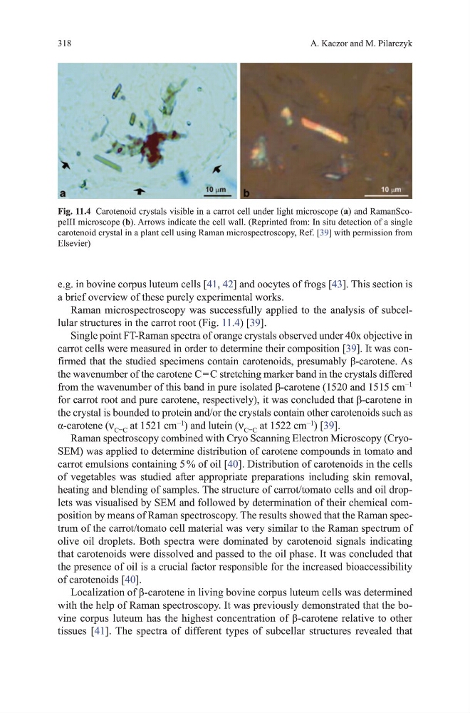
Raman microspectroscopy was successfully applied to the analysis of subcellular structures in the carrot root (Fig. 11.4) [39].
Single point Ft-Raman spectra of orange crystals observed under 40x objective in
carrot cells were measured in order to determine their composition [39]. It was confirmed that the studied specimens contain carotenoids, presumably β-carotene. As
the wavenumber of the carotene C = C stretching marker band in the crystals differed
from the wavenumber of this band in pure isolated β-carotene (1520 and 1515 cm
−1
for carrot root and pure carotene, respectively), it was concluded that β-carotene in
the crystal is bounded to protein and/or the crystals contain other carotenoids such as
α-carotene (ν C=C at 1521 cm
−1
) and lutein (ν C=C at 1522 cm
−1
) [39].
Raman spectroscopy combined with Cryo Scanning Electron microscopy (CryoSEm) was applied to determine distribution of carotene compounds in tomato and
carrot emulsions containing 5 % of oil [40]. distribution of carotenoids in the cells
of vegetables was studied after appropriate preparations including skin removal,
heating and blending of samples. the structure of carrot/tomato cells and oil droplets was visualised by SEm and followed by determination of their chemical composition by means of Raman spectroscopy. the results showed that the Raman spectrum of the carrot/tomato cell material was very similar to the Raman spectrum of
olive oil droplets. Both spectra were dominated by carotenoid signals indicating
that carotenoids were dissolved and passed to the oil phase. It was concluded that
the presence of oil is a crucial factor responsible for the increased bioaccessibility
of carotenoids [40].
Localization of β-carotene in living bovine corpus luteum cells was determined
with the help of Raman spectroscopy. It was previously demonstrated that the bovine corpus luteum has the highest concentration of β-carotene relative to other
tissues [41]. the spectra of different types of subcellar structures revealed that
A. Kaczor and m. Pilarczyk
Fig. 11.4 Carotenoid crystals visible in a carrot cell under light microscope (a) and RamanScopeIII microscope (b). Arrows indicate the cell wall. (Reprinted from: In situ detection of a single
carotenoid crystal in a plant cell using Raman microspectroscopy, Ref. [39] with permission from
Elsevier)
e.g. in bovine corpus luteum cells [41, 42] and oocytes of frogs [43]. this section is
a brief overview of these purely experimental works.
Raman microspectroscopy was successfully applied to the analysis of subcellular structures in the carrot root (Fig. 11.4) [39].
Single point Ft-Raman spectra of orange crystals observed under 40x objective in
carrot cells were measured in order to determine their composition [39]. It was confirmed that the studied specimens contain carotenoids, presumably β-carotene. As
the wavenumber of the carotene C = C stretching marker band in the crystals differed
from the wavenumber of this band in pure isolated β-carotene (1520 and 1515 cm
−1
for carrot root and pure carotene, respectively), it was concluded that β-carotene in
the crystal is bounded to protein and/or the crystals contain other carotenoids such as
α-carotene (ν C=C at 1521 cm
−1
) and lutein (ν C=C at 1522 cm
−1
) [39].
Raman spectroscopy combined with Cryo Scanning Electron microscopy (CryoSEm) was applied to determine distribution of carotene compounds in tomato and
carrot emulsions containing 5 % of oil [40]. distribution of carotenoids in the cells
of vegetables was studied after appropriate preparations including skin removal,
heating and blending of samples. the structure of carrot/tomato cells and oil droplets was visualised by SEm and followed by determination of their chemical composition by means of Raman spectroscopy. the results showed that the Raman spectrum of the carrot/tomato cell material was very similar to the Raman spectrum of
olive oil droplets. Both spectra were dominated by carotenoid signals indicating
that carotenoids were dissolved and passed to the oil phase. It was concluded that
the presence of oil is a crucial factor responsible for the increased bioaccessibility
of carotenoids [40].
Localization of β-carotene in living bovine corpus luteum cells was determined
with the help of Raman spectroscopy. It was previously demonstrated that the bovine corpus luteum has the highest concentration of β-carotene relative to other
tissues [41]. the spectra of different types of subcellar structures revealed that
A. Kaczor and m. Pilarczyk
Fig. 11.4 Carotenoid crystals visible in a carrot cell under light microscope (a) and RamanScopeIII microscope (b). Arrows indicate the cell wall. (Reprinted from: In situ detection of a single
carotenoid crystal in a plant cell using Raman microspectroscopy, Ref. [39] with permission from
Elsevier)