

10 Chemometric Analysis of Raman and IR Spectra of Natural Dyes
301
10.3.3 Anthraquinones
In spite of the fact that anthraquinones are widely distributed pigments in nature
[6], the vibrational studies of them are not so popular like for the other colorants.
the pigments founded in lichens, parietin [85, 104–106] and emodin [104] were
detected in natural samples, but quantification analysis was carried out only for
parietin accompanied by carotenoids [85]. generally, the analysis were made with
the NIR laser excitation wavelength (1,064 nm).
most papers about the detection of anthraquinones shows an application of a
SERS technique to in situ analysis. however, most of them are related to the determination of dye/pigment on the thread in context of the conservation of art [31, 34,
107–109], but not in the biological systems.
10.3.4 Indigo
Indigo shows extremely intense signals in its Raman spectrum that enables to detect
this dye directly in textiles and plants [41, 110].
one of the plant species, which contains the indigo precursor, indican, is a dyer’s
knotweed ( Polygonum tinctorium). In enzymatically catalyzed process, indigo is
formed from indican upon leaf drying and in the case of leaf surface damaging.
Both these processes were observed with Ft-Raman spectroscopy during two days
of drying at room temperature (Fig. 10.15) [41]. the blue area on the leaf surface
indicates damages of the plant tissue where the indican was enzymatically transformed to the indigo. the maps were obtained by integration of the marker band
near 1,570 cm
−1
. It is clearly seen that firstly the indigo is present only in the damaged area of a fresh leaf, whilst upon two days, the amount of indigo gradually
increased in the leaf area.
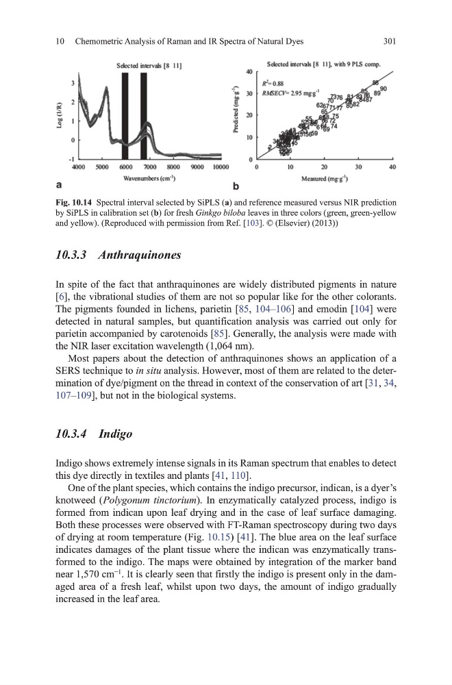
Fig. 10.14 Spectral interval selected by SiPLS (a) and reference measured versus NIR prediction
by SiPLS in calibration set (b) for fresh Ginkgo biloba leaves in three colors ( green, green-yellow
and yellow). (Reproduced with permission from Ref. [103]. © (Elsevier) (2013))
301
10.3.3 Anthraquinones
In spite of the fact that anthraquinones are widely distributed pigments in nature
[6], the vibrational studies of them are not so popular like for the other colorants.
the pigments founded in lichens, parietin [85, 104–106] and emodin [104] were
detected in natural samples, but quantification analysis was carried out only for
parietin accompanied by carotenoids [85]. generally, the analysis were made with
the NIR laser excitation wavelength (1,064 nm).
most papers about the detection of anthraquinones shows an application of a
SERS technique to in situ analysis. however, most of them are related to the determination of dye/pigment on the thread in context of the conservation of art [31, 34,
107–109], but not in the biological systems.
10.3.4 Indigo
Indigo shows extremely intense signals in its Raman spectrum that enables to detect
this dye directly in textiles and plants [41, 110].
one of the plant species, which contains the indigo precursor, indican, is a dyer’s
knotweed ( Polygonum tinctorium). In enzymatically catalyzed process, indigo is
formed from indican upon leaf drying and in the case of leaf surface damaging.
Both these processes were observed with Ft-Raman spectroscopy during two days
of drying at room temperature (Fig. 10.15) [41]. the blue area on the leaf surface
indicates damages of the plant tissue where the indican was enzymatically transformed to the indigo. the maps were obtained by integration of the marker band
near 1,570 cm
−1
. It is clearly seen that firstly the indigo is present only in the damaged area of a fresh leaf, whilst upon two days, the amount of indigo gradually
increased in the leaf area.
Fig. 10.14 Spectral interval selected by SiPLS (a) and reference measured versus NIR prediction
by SiPLS in calibration set (b) for fresh Ginkgo biloba leaves in three colors ( green, green-yellow
and yellow). (Reproduced with permission from Ref. [103]. © (Elsevier) (2013))