

164
M. Pecul and W. Dzwolak
means of this model [20, 35, 36, 38, 46]. other chromophores contributing to the
Cd spectrum of peptides and proteins are aromatic groups. For them, the transition
dipoles are nearly additive, and they are very suitable for calculations using the
exciton coupling model. Since they can provide unique structural information (see
below), they have been the subject of a number of computational studies [20, 47,
48]. the Cd spectrum of the disulphide chromophore, although less important in
practice, has also been modeled [29, 49, 50].
6.3 Applications of CD in Proteomics
the ubiquitous left-handedness of all except one (glycine) translational amino acids,
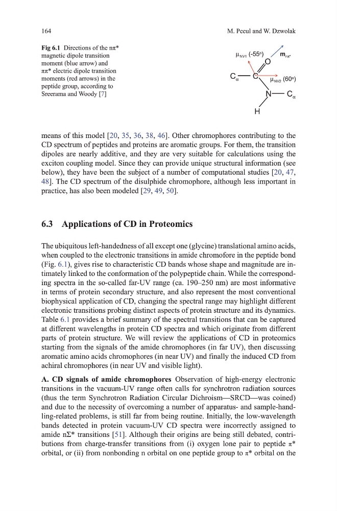
when coupled to the electronic transitions in amide chromofore in the peptide bond
(Fig. 6.1), gives rise to characteristic Cd bands whose shape and magnitude are intimately linked to the conformation of the polypeptide chain. While the corresponding spectra in the so-called far-uv range (ca. 190–250 nm) are most informative
in terms of protein secondary structure, and also represent the most conventional
biophysical application of Cd, changing the spectral range may highlight different
electronic transitions probing distinct aspects of protein structure and its dynamics.
table 6.1 provides a brief summary of the spectral transitions that can be captured
at different wavelengths in protein Cd spectra and which originate from different
parts of protein structure. We will review the applications of Cd in proteomics
starting from the signals of the amide chromophores (in far uv), then discussing
aromatic amino acids chromophores (in near uv) and finally the induced Cd from
achiral chromophores (in near uv and visible light).
A. CD signals of amide chromophores observation of high-energy electronic
transitions in the vacuum-uv range often calls for synchrotron radiation sources
(thus the term Synchrotron Radiation Circular dichroism—SRCd—was coined)
and due to the necessity of overcoming a number of apparatus- and sample-handling-related problems, is still far from being routine. Initially, the low-wavelength
bands detected in protein vacuum-uv Cd spectra were incorrectly assigned to
amide nΣ* transitions [51]. Although their origins are being still debated, contributions from charge-transfer transitions from (i) oxygen lone pair to peptide π*
orbital, or (ii) from nonbonding n orbital on one peptide group to π* orbital on the
α
α
π
µ
µ
Fig 6.1 Directions of the nπ*
magnetic dipole transition
moment (blue arrow) and
ππ* electric dipole transition
moments (red arrows) in the
peptide group, according to
Sreerama and Woody [7]
M. Pecul and W. Dzwolak
means of this model [20, 35, 36, 38, 46]. other chromophores contributing to the
Cd spectrum of peptides and proteins are aromatic groups. For them, the transition
dipoles are nearly additive, and they are very suitable for calculations using the
exciton coupling model. Since they can provide unique structural information (see
below), they have been the subject of a number of computational studies [20, 47,
48]. the Cd spectrum of the disulphide chromophore, although less important in
practice, has also been modeled [29, 49, 50].
6.3 Applications of CD in Proteomics
the ubiquitous left-handedness of all except one (glycine) translational amino acids,
when coupled to the electronic transitions in amide chromofore in the peptide bond
(Fig. 6.1), gives rise to characteristic Cd bands whose shape and magnitude are intimately linked to the conformation of the polypeptide chain. While the corresponding spectra in the so-called far-uv range (ca. 190–250 nm) are most informative
in terms of protein secondary structure, and also represent the most conventional
biophysical application of Cd, changing the spectral range may highlight different
electronic transitions probing distinct aspects of protein structure and its dynamics.
table 6.1 provides a brief summary of the spectral transitions that can be captured
at different wavelengths in protein Cd spectra and which originate from different
parts of protein structure. We will review the applications of Cd in proteomics
starting from the signals of the amide chromophores (in far uv), then discussing
aromatic amino acids chromophores (in near uv) and finally the induced Cd from
achiral chromophores (in near uv and visible light).
A. CD signals of amide chromophores observation of high-energy electronic
transitions in the vacuum-uv range often calls for synchrotron radiation sources
(thus the term Synchrotron Radiation Circular dichroism—SRCd—was coined)
and due to the necessity of overcoming a number of apparatus- and sample-handling-related problems, is still far from being routine. Initially, the low-wavelength
bands detected in protein vacuum-uv Cd spectra were incorrectly assigned to
amide nΣ* transitions [51]. Although their origins are being still debated, contributions from charge-transfer transitions from (i) oxygen lone pair to peptide π*
orbital, or (ii) from nonbonding n orbital on one peptide group to π* orbital on the
α
α
π
µ
µ
Fig 6.1 Directions of the nπ*
magnetic dipole transition
moment (blue arrow) and
ππ* electric dipole transition
moments (red arrows) in the
peptide group, according to
Sreerama and Woody [7]