

5 α-Amino Acids In Water: A Review of VCD and ROA Spectra
131
5.5.2.7 L-methionine
O
OH
NH 2
S
C
H 3
Scheme 10 (S)-2-amino(methylthio) butanoic acid
VCD and ROA: Experiment and Calculations So far, for L-methionine, the only
vCd measurements have been done by the Nafie group in the 1980s [147–150],
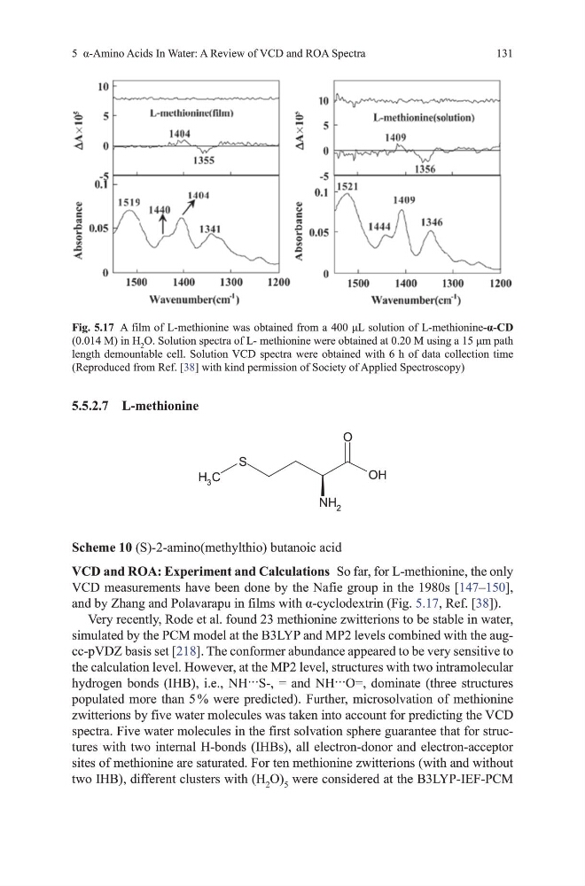
and by Zhang and Polavarapu in films with α-cyclodextrin (Fig. 5.17, Ref. [38]).
very recently, Rode et al. found 23 methionine zwitterions to be stable in water,
simulated by the PCm model at the B3LYP and mP2 levels combined with the augcc-pvdZ basis set [218]. the conformer abundance appeared to be very sensitive to
the calculation level. however, at the mP2 level, structures with two intramolecular
hydrogen bonds (IHB), i.e., NH···S-, = and NH···O=, dominate (three structures
populated more than 5 % were predicted). Further, microsolvation of methionine
zwitterions by five water molecules was taken into account for predicting the vCd
spectra. Five water molecules in the first solvation sphere guarantee that for structures with two internal h-bonds (IhBs), all electron-donor and electron-acceptor
sites of methionine are saturated. For ten methionine zwitterions (with and without
two IhB), different clusters with (h 2 o) 5 were considered at the B3LYP-IEF-PCm
Fig. 5.17 A film of L-methionine was obtained from a 400 μL solution of L-methionine-α-CD
(0.014 m) in h 2 O. Solution spectra of L- methionine were obtained at 0.20 M using a 15 μm path
length demountable cell. Solution vCd spectra were obtained with 6 h of data collection time
(Reproduced from Ref. [38] with kind permission of Society of Applied Spectroscopy)
131
5.5.2.7 L-methionine
O
OH
NH 2
S
C
H 3
Scheme 10 (S)-2-amino(methylthio) butanoic acid
VCD and ROA: Experiment and Calculations So far, for L-methionine, the only
vCd measurements have been done by the Nafie group in the 1980s [147–150],
and by Zhang and Polavarapu in films with α-cyclodextrin (Fig. 5.17, Ref. [38]).
very recently, Rode et al. found 23 methionine zwitterions to be stable in water,
simulated by the PCm model at the B3LYP and mP2 levels combined with the augcc-pvdZ basis set [218]. the conformer abundance appeared to be very sensitive to
the calculation level. however, at the mP2 level, structures with two intramolecular
hydrogen bonds (IHB), i.e., NH···S-, = and NH···O=, dominate (three structures
populated more than 5 % were predicted). Further, microsolvation of methionine
zwitterions by five water molecules was taken into account for predicting the vCd
spectra. Five water molecules in the first solvation sphere guarantee that for structures with two internal h-bonds (IhBs), all electron-donor and electron-acceptor
sites of methionine are saturated. For ten methionine zwitterions (with and without
two IhB), different clusters with (h 2 o) 5 were considered at the B3LYP-IEF-PCm
Fig. 5.17 A film of L-methionine was obtained from a 400 μL solution of L-methionine-α-CD
(0.014 m) in h 2 O. Solution spectra of L- methionine were obtained at 0.20 M using a 15 μm path
length demountable cell. Solution vCd spectra were obtained with 6 h of data collection time
(Reproduced from Ref. [38] with kind permission of Society of Applied Spectroscopy)