

5 α-Amino Acids In Water: A Review of VCD and ROA Spectra
123
the predominant contributions of the electric dipole–magnetic dipole invariant to
the RoA signals in L-proline.
Finally, the RoA spectra of 4( R)-hydroxyproline in aqueous solutions at three
different ph values were interpreted by B3LYP/IEF-PCm/aug-cc-pvdZ calculations for 10 anionic, 5 cationic and 15 zwitterionic conformations (Fig. 5.11)
[208]. the RoA intensities were calculated at the hF/aug-ccpvdZ level, for
which using the polarisable continuum model for calculations of optical tensors
was found to be particularly problematic for the zwitterionic structures. As for
proline, the endo forms of hydroxyproline appeared to be the most stable conformers. the RoA spectra calculated for the zwitterionic and cation forms stem
predominantly from the two lowest conformations each, whereas for the isolated
anion, the spectrum originated from one while in aqueous solution from four lowlying conformers. the largest RoA intensities, negative or positive, were assigned
to stretching vibrations of the five-membered ring of 4-trans-(R)-hydroxyproline.
No simple correlation between the ring conformation and the RoA intensity of
those vibrations was found.
5.5.2.3 L-cysteine
O
OH
NH 2
S
H
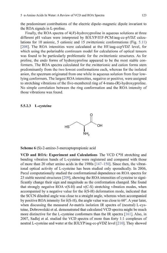
Scheme 6 (S)-2-amino-3-mercaptopropionic acid
VCD and ROA: Experiment and Calculations the vCd C*h stretching and
bending vibration bands of L-cysteine were registered and compared with those
of more than 20 other amino acids in the 1980s [147–150]. Since then, the vibrational optical activity of L-cysteine has been studied only sporadically. In 2006,
Pecul computationally studied the conformational dependence on RoA spectra for
23 stable neutral structures [209], showing the RoA intensities of cysteine to significantly change their sign and magnitude as the conformation changed. She found
that strongly negative ROA ν(S-H) and ν(C-S) stretching vibration modes, when
accompanied by a negative value for the δ(S-H) deformation mode, indicated that
the SCCN dihedral angle was close to a straight angle, whereas when accompanied
by positive ROA intensity for δ(S-H), the angle value was close to 60°. A year later,
when discussing the measured Ar-matrix isolation IR spectra of (neutral) L-cysteine, dobrowolski et al. demonstrated that calculated vCd spectra might be much
more distinctive for the L-cysteine conformers than the IR spectra [161]. Also, in
2007, Sadlej et al. studied the vCd spectra of more than forty 1:1 complexes of
neutral L-cysteine and water at the B3LYP/aug-cc-pvdZ level [210]. they showed
123
the predominant contributions of the electric dipole–magnetic dipole invariant to
the RoA signals in L-proline.
Finally, the RoA spectra of 4( R)-hydroxyproline in aqueous solutions at three
different ph values were interpreted by B3LYP/IEF-PCm/aug-cc-pvdZ calculations for 10 anionic, 5 cationic and 15 zwitterionic conformations (Fig. 5.11)
[208]. the RoA intensities were calculated at the hF/aug-ccpvdZ level, for
which using the polarisable continuum model for calculations of optical tensors
was found to be particularly problematic for the zwitterionic structures. As for
proline, the endo forms of hydroxyproline appeared to be the most stable conformers. the RoA spectra calculated for the zwitterionic and cation forms stem
predominantly from the two lowest conformations each, whereas for the isolated
anion, the spectrum originated from one while in aqueous solution from four lowlying conformers. the largest RoA intensities, negative or positive, were assigned
to stretching vibrations of the five-membered ring of 4-trans-(R)-hydroxyproline.
No simple correlation between the ring conformation and the RoA intensity of
those vibrations was found.
5.5.2.3 L-cysteine
O
OH
NH 2
S
H
Scheme 6 (S)-2-amino-3-mercaptopropionic acid
VCD and ROA: Experiment and Calculations the vCd C*h stretching and
bending vibration bands of L-cysteine were registered and compared with those
of more than 20 other amino acids in the 1980s [147–150]. Since then, the vibrational optical activity of L-cysteine has been studied only sporadically. In 2006,
Pecul computationally studied the conformational dependence on RoA spectra for
23 stable neutral structures [209], showing the RoA intensities of cysteine to significantly change their sign and magnitude as the conformation changed. She found
that strongly negative ROA ν(S-H) and ν(C-S) stretching vibration modes, when
accompanied by a negative value for the δ(S-H) deformation mode, indicated that
the SCCN dihedral angle was close to a straight angle, whereas when accompanied
by positive ROA intensity for δ(S-H), the angle value was close to 60°. A year later,
when discussing the measured Ar-matrix isolation IR spectra of (neutral) L-cysteine, dobrowolski et al. demonstrated that calculated vCd spectra might be much
more distinctive for the L-cysteine conformers than the IR spectra [161]. Also, in
2007, Sadlej et al. studied the vCd spectra of more than forty 1:1 complexes of
neutral L-cysteine and water at the B3LYP/aug-cc-pvdZ level [210]. they showed