

which have absorbed lower wavelengths are more likely to dissociate within the
measurement time window, see Fig. 3.7.
3.1.3 Reflectron Time-of-Flight Mass Spectrometer
Another single pass instrument that can be used to study photofragmentation is a
reflectron time-of-flight (R-TOF) mass spectrometer (Fig. 3.16) [24]. In the
depicted instrument, ions are produced using electrospray, desolvated in a heated
capillary, and accumulated in a hexapole ion trap. They are subsequently extracted,
focused and injected into the acceleration region of a TOF mass spectrometer.
There ions are accelerated perpendicularly into a flight tube, and those of interest
irradiated using a nanosecond-pulsed tuneable laser. A two-stage reflectron is then
used to separate ions according to their mass-to-charge ratio, and the ions are
detected on a microchannel plate detector. A conversion dynode and channeltron
located next to the TOF acceleration region can be used for ion beam diagnostics.
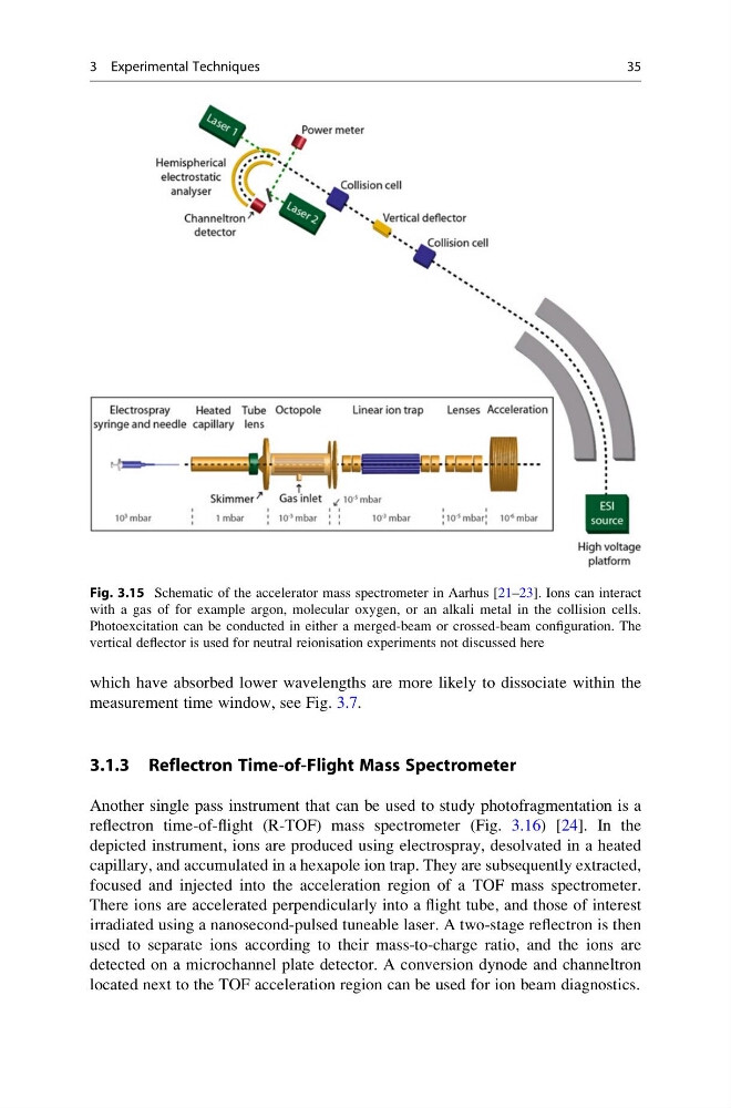
Fig. 3.15 Schematic of the accelerator mass spectrometer in Aarhus [21–23]. Ions can interact
with a gas of for example argon, molecular oxygen, or an alkali metal in the collision cells.
Photoexcitation can be conducted in either a merged-beam or crossed-beam configuration. The
vertical deflector is used for neutral reionisation experiments not discussed here
3 Experimental Techniques
35
measurement time window, see Fig. 3.7.
3.1.3 Reflectron Time-of-Flight Mass Spectrometer
Another single pass instrument that can be used to study photofragmentation is a
reflectron time-of-flight (R-TOF) mass spectrometer (Fig. 3.16) [24]. In the
depicted instrument, ions are produced using electrospray, desolvated in a heated
capillary, and accumulated in a hexapole ion trap. They are subsequently extracted,
focused and injected into the acceleration region of a TOF mass spectrometer.
There ions are accelerated perpendicularly into a flight tube, and those of interest
irradiated using a nanosecond-pulsed tuneable laser. A two-stage reflectron is then
used to separate ions according to their mass-to-charge ratio, and the ions are
detected on a microchannel plate detector. A conversion dynode and channeltron
located next to the TOF acceleration region can be used for ion beam diagnostics.
Fig. 3.15 Schematic of the accelerator mass spectrometer in Aarhus [21–23]. Ions can interact
with a gas of for example argon, molecular oxygen, or an alkali metal in the collision cells.
Photoexcitation can be conducted in either a merged-beam or crossed-beam configuration. The
vertical deflector is used for neutral reionisation experiments not discussed here
3 Experimental Techniques
35