

Nearby electric fields set up by an environment will perturb the electronic
structure of the chromophore and influence the transition energies, dependent on
the character of the electronic transition, and particularly the degree of charge
transfer (CT) (Fig. 2.3). Positive or negative charges close to the chromophore
can for example originate from acidic or basic amino acid residues (Figs. 2.4 and
2.5). To elucidate electric-field effects and whether an environment is innocent or
not, it is useful (or even necessary) to know the intrinsic absorption spectrum of the
isolated chromophore!
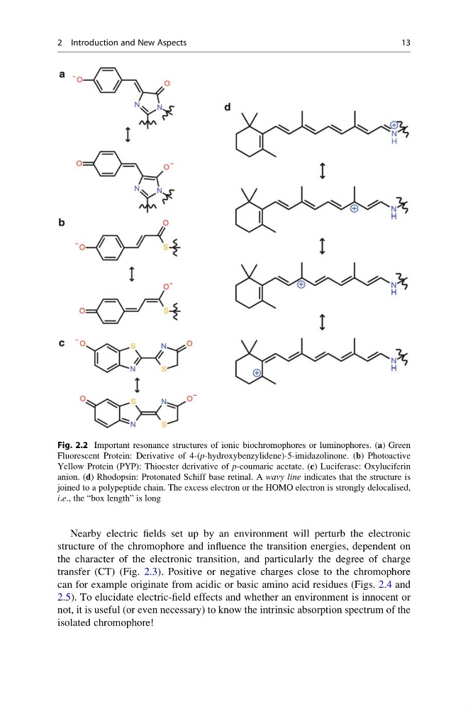
Fig. 2.2 Important resonance structures of ionic biochromophores or luminophores. (a) Green
Fluorescent Protein: Derivative of 4-(p-hydroxybenzylidene)-5-imidazolinone. (b) Photoactive
Yellow Protein (PYP): Thioester derivative of p-coumaric acetate. (c) Luciferase: Oxyluciferin
anion. (d) Rhodopsin: Protonated Schiff base retinal. A wavy line indicates that the structure is
joined to a polypeptide chain. The excess electron or the HOMO electron is strongly delocalised,
i.e., the “box length” is long
2 Introduction and New Aspects
13
structure of the chromophore and influence the transition energies, dependent on
the character of the electronic transition, and particularly the degree of charge
transfer (CT) (Fig. 2.3). Positive or negative charges close to the chromophore
can for example originate from acidic or basic amino acid residues (Figs. 2.4 and
2.5). To elucidate electric-field effects and whether an environment is innocent or
not, it is useful (or even necessary) to know the intrinsic absorption spectrum of the
isolated chromophore!
Fig. 2.2 Important resonance structures of ionic biochromophores or luminophores. (a) Green
Fluorescent Protein: Derivative of 4-(p-hydroxybenzylidene)-5-imidazolinone. (b) Photoactive
Yellow Protein (PYP): Thioester derivative of p-coumaric acetate. (c) Luciferase: Oxyluciferin
anion. (d) Rhodopsin: Protonated Schiff base retinal. A wavy line indicates that the structure is
joined to a polypeptide chain. The excess electron or the HOMO electron is strongly delocalised,
i.e., the “box length” is long
2 Introduction and New Aspects
13