

state levels. This simple approach benefits from providing a direct estimate of the
excitation energy but it suffers from not considering electron–electron interactions,
and also the spacing between the energy levels increases with the quantum number
instead of decreasing as they duly should. A chemist will use molecular orbital
theory, and from linear combinations of atomic orbitals, the Aufbau principle and
the Pauli principle reach the same conclusion regarding the intricate connection
between spatial delocalisation and excitation energy based on the number of
alternating double and single bonds. Here the qualitative picture is correct but a
quick calculation of excitation energy is difficult, if not impossible. Examples of
highly π-conjugated biochromophores include the aromatic amino acids (phenylalanine, tyrosine, and tryptophan), the nucleic acid bases (adenine, thymine, guanine,
cytosine, and uracil), porphyrins and metalloporphyrins (e.g., chlorophylls and
heme), and several protein biochromophores responsible for example for vision,
light emission, and signalling. The latter ones absorb in the visible while the former
ones (aromatic amino acids and bases) absorb in the UV. Structures of some of the
chromophores can be found here, in the other chapters, and in the concepts pages.
Typical ionic chromophores like the anions within the Green Fluorescent Protein
(GFP) (the topic of Chap. 5 by Andersen and Bochenkova) and Photoactive Yellow
Protein (PYP), the oxyluciferin anion located in the luciferase enzyme and responsible for firefly light emission, and the protonated Schiff base retinal within the
rhodopsin vision protein absorb (or emit) in the visible [1]. These are buried within
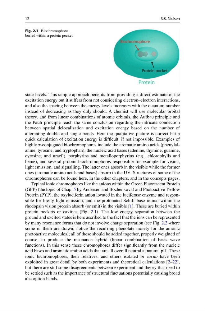
protein pockets or cavities (Fig. 2.1). The low energy separation between the
ground and excited states is here ascribed to the fact that the ions can be represented
by many resonance forms that do not involve charge separation (see Fig. 2.2 where
some of them are drawn; notice the recurring phenolate moiety for the anionic
photoactive molecules); all of these should be added together, properly weighted of
course, to produce the resonance hybrid (linear combination of basis wave
functions). In this sense these chromophores differ significantly from the nucleic
acid bases and aromatic amino acids that are all overall neutral at natural pH. These
ionic bichromophores, their relatives, and others isolated in vacuo have been
exploited in great detail by both experiments and theoretical calculations [2–22],
but there are still some disagreements between experiment and theory that need to
be settled such as the importance of structural fluctuations potentially causing broad
absorption bands.
Fig. 2.1 Biochromophore
buried within a protein pocket
12
S.B. Nielsen
excitation energy but it suffers from not considering electron–electron interactions,
and also the spacing between the energy levels increases with the quantum number
instead of decreasing as they duly should. A chemist will use molecular orbital
theory, and from linear combinations of atomic orbitals, the Aufbau principle and
the Pauli principle reach the same conclusion regarding the intricate connection
between spatial delocalisation and excitation energy based on the number of
alternating double and single bonds. Here the qualitative picture is correct but a
quick calculation of excitation energy is difficult, if not impossible. Examples of
highly π-conjugated biochromophores include the aromatic amino acids (phenylalanine, tyrosine, and tryptophan), the nucleic acid bases (adenine, thymine, guanine,
cytosine, and uracil), porphyrins and metalloporphyrins (e.g., chlorophylls and
heme), and several protein biochromophores responsible for example for vision,
light emission, and signalling. The latter ones absorb in the visible while the former
ones (aromatic amino acids and bases) absorb in the UV. Structures of some of the
chromophores can be found here, in the other chapters, and in the concepts pages.
Typical ionic chromophores like the anions within the Green Fluorescent Protein
(GFP) (the topic of Chap. 5 by Andersen and Bochenkova) and Photoactive Yellow
Protein (PYP), the oxyluciferin anion located in the luciferase enzyme and responsible for firefly light emission, and the protonated Schiff base retinal within the
rhodopsin vision protein absorb (or emit) in the visible [1]. These are buried within
protein pockets or cavities (Fig. 2.1). The low energy separation between the
ground and excited states is here ascribed to the fact that the ions can be represented
by many resonance forms that do not involve charge separation (see Fig. 2.2 where
some of them are drawn; notice the recurring phenolate moiety for the anionic
photoactive molecules); all of these should be added together, properly weighted of
course, to produce the resonance hybrid (linear combination of basis wave
functions). In this sense these chromophores differ significantly from the nucleic
acid bases and aromatic amino acids that are all overall neutral at natural pH. These
ionic bichromophores, their relatives, and others isolated in vacuo have been
exploited in great detail by both experiments and theoretical calculations [2–22],
but there are still some disagreements between experiment and theory that need to
be settled such as the importance of structural fluctuations potentially causing broad
absorption bands.
Fig. 2.1 Biochromophore
buried within a protein pocket
12
S.B. Nielsen