

11.4 Soft X-Ray Photofragmentation of Protonated
Polypeptides
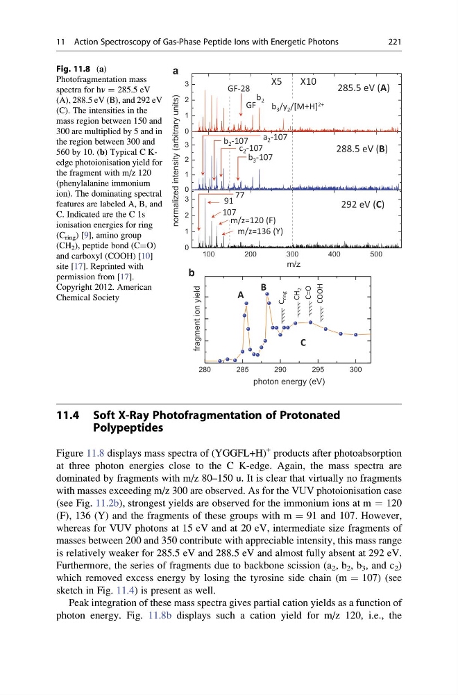
Figure 11.8 displays mass spectra of (YGGFL+H)
+ products after photoabsorption
at three photon energies close to the C K-edge. Again, the mass spectra are
dominated by fragments with m/z 80–150 u. It is clear that virtually no fragments
with masses exceeding m/z 300 are observed. As for the VUV photoionisation case
(see Fig. 11.2b), strongest yields are observed for the immonium ions at m ¼ 120
(F), 136 (Y) and the fragments of these groups with m ¼ 91 and 107. However,
whereas for VUV photons at 15 eV and at 20 eV, intermediate size fragments of
masses between 200 and 350 contribute with appreciable intensity, this mass range
is relatively weaker for 285.5 eV and 288.5 eV and almost fully absent at 292 eV.
Furthermore, the series of fragments due to backbone scission (a 2 , b 2 , b 3 , and c 2 )
which removed excess energy by losing the tyrosine side chain (m ¼ 107) (see
sketch in Fig. 11.4) is present as well.
Peak integration of these mass spectra gives partial cation yields as a function of
photon energy. Fig. 11.8b displays such a cation yield for m/z 120, i.e., the
100
200
300
400
500
0
1
2
3
0
1
2
3
0
1
2
3
m/z
280
285
290
295
300
fragment ion yield
photon energy (eV)
C=O
COOH
CH
2
C
ring
A
B
C
285.5 eV (A)
288.5 eV (B)
292 eV (C)
a
b
m/z=136 (Y)
107
91
GF
b 2
b 3 /y 2 /[M+H] 2+
GF-28
X5
X10
m/z=120 (F)
b 2 -107
b 3 -107
c 2 -107
77
a 2 -107
normalized intensity (arbitrary units)
Fig. 11.8 (a)
Photofragmentation mass
spectra for hν ¼ 285.5 eV
(A), 288.5 eV (B), and 292 eV
(C). The intensities in the
mass region between 150 and
300 are multiplied by 5 and in
the region between 300 and
560 by 10. (b) Typical C Kedge photoionisation yield for
the fragment with m/z 120
(phenylalanine immonium
ion). The dominating spectral
features are labeled A, B, and
C. Indicated are the C 1s
ionisation energies for ring
(C ring ) [9], amino group
(CH 2 ), peptide bond (C¼O)
and carboxyl (COOH) [10]
site [17]. Reprinted with
permission from [17].
Copyright 2012. American
Chemical Society
11 Action Spectroscopy of Gas-Phase Peptide Ions with Energetic Photons
221
Polypeptides
Figure 11.8 displays mass spectra of (YGGFL+H)
+ products after photoabsorption
at three photon energies close to the C K-edge. Again, the mass spectra are
dominated by fragments with m/z 80–150 u. It is clear that virtually no fragments
with masses exceeding m/z 300 are observed. As for the VUV photoionisation case
(see Fig. 11.2b), strongest yields are observed for the immonium ions at m ¼ 120
(F), 136 (Y) and the fragments of these groups with m ¼ 91 and 107. However,
whereas for VUV photons at 15 eV and at 20 eV, intermediate size fragments of
masses between 200 and 350 contribute with appreciable intensity, this mass range
is relatively weaker for 285.5 eV and 288.5 eV and almost fully absent at 292 eV.
Furthermore, the series of fragments due to backbone scission (a 2 , b 2 , b 3 , and c 2 )
which removed excess energy by losing the tyrosine side chain (m ¼ 107) (see
sketch in Fig. 11.4) is present as well.
Peak integration of these mass spectra gives partial cation yields as a function of
photon energy. Fig. 11.8b displays such a cation yield for m/z 120, i.e., the
100
200
300
400
500
0
1
2
3
0
1
2
3
0
1
2
3
m/z
280
285
290
295
300
fragment ion yield
photon energy (eV)
C=O
COOH
CH
2
C
ring
A
B
C
285.5 eV (A)
288.5 eV (B)
292 eV (C)
a
b
m/z=136 (Y)
107
91
GF
b 2
b 3 /y 2 /[M+H] 2+
GF-28
X5
X10
m/z=120 (F)
b 2 -107
b 3 -107
c 2 -107
77
a 2 -107
normalized intensity (arbitrary units)
Fig. 11.8 (a)
Photofragmentation mass
spectra for hν ¼ 285.5 eV
(A), 288.5 eV (B), and 292 eV
(C). The intensities in the
mass region between 150 and
300 are multiplied by 5 and in
the region between 300 and
560 by 10. (b) Typical C Kedge photoionisation yield for
the fragment with m/z 120
(phenylalanine immonium
ion). The dominating spectral
features are labeled A, B, and
C. Indicated are the C 1s
ionisation energies for ring
(C ring ) [9], amino group
(CH 2 ), peptide bond (C¼O)
and carboxyl (COOH) [10]
site [17]. Reprinted with
permission from [17].
Copyright 2012. American
Chemical Society
11 Action Spectroscopy of Gas-Phase Peptide Ions with Energetic Photons
221