

glycine moieties allows controlled variation of photoabsorption cross section and
charge/energy migration distance. Figure 11.6 shows a comparison of spectra
obtained at hν ¼ 20 eV for n ¼ 0, 1, 3, 5, and 10. The mass of the protonated
peptides increases from 329 to 899, which is why the mass range is increasing. For
all peptides except [YG 10 F+H]
+ fragments related to the aromatic sidechains are
almost exclusively observed. Even though the fragment distribution strongly varies
with n, in all cases the spectra are dominated by masses smaller than 200, as found
for leucine enkephalin. These fragments all stem from the two termini, indicating
that not only photoabsorption in an aromatic sidechain leads to formation of the
respective ion. Rather, the same fragments can also be produced upon
photoabsorption by the peptide backbone, possibly after fast charge or energy
transfer towards the termini.
Y sidechain ionizaƟon
fast Y sidechain caƟon loss
backbone cleavage aŌer IVR
a
c 2
c 3
b 2
b 4
a 4
b 3
c
b
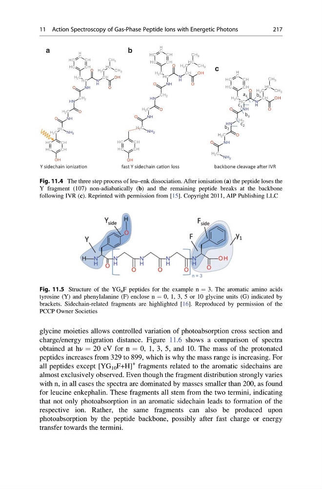
Fig. 11.4 The three step process of leu–enk dissociation. After ionisation (a) the peptide loses the
Y fragment (107) non-adiabatically (b) and the remaining peptide breaks at the backbone
following IVR (c). Reprinted with permission from [15]. Copyright 2011, AIP Publishing LLC
Fig. 11.5 Structure of the YG n F peptides for the example n ¼ 3. The aromatic amino acids
tyrosine (Y) and phenylalanine (F) enclose n ¼ 0, 1, 3, 5 or 10 glycine units (G) indicated by
brackets. Sidechain-related fragments are highlighted [16]. Reproduced by permission of the
PCCP Owner Societies
11 Action Spectroscopy of Gas-Phase Peptide Ions with Energetic Photons
217
charge/energy migration distance. Figure 11.6 shows a comparison of spectra
obtained at hν ¼ 20 eV for n ¼ 0, 1, 3, 5, and 10. The mass of the protonated
peptides increases from 329 to 899, which is why the mass range is increasing. For
all peptides except [YG 10 F+H]
+ fragments related to the aromatic sidechains are
almost exclusively observed. Even though the fragment distribution strongly varies
with n, in all cases the spectra are dominated by masses smaller than 200, as found
for leucine enkephalin. These fragments all stem from the two termini, indicating
that not only photoabsorption in an aromatic sidechain leads to formation of the
respective ion. Rather, the same fragments can also be produced upon
photoabsorption by the peptide backbone, possibly after fast charge or energy
transfer towards the termini.
Y sidechain ionizaƟon
fast Y sidechain caƟon loss
backbone cleavage aŌer IVR
a
c 2
c 3
b 2
b 4
a 4
b 3
c
b
Fig. 11.4 The three step process of leu–enk dissociation. After ionisation (a) the peptide loses the
Y fragment (107) non-adiabatically (b) and the remaining peptide breaks at the backbone
following IVR (c). Reprinted with permission from [15]. Copyright 2011, AIP Publishing LLC
Fig. 11.5 Structure of the YG n F peptides for the example n ¼ 3. The aromatic amino acids
tyrosine (Y) and phenylalanine (F) enclose n ¼ 0, 1, 3, 5 or 10 glycine units (G) indicated by
brackets. Sidechain-related fragments are highlighted [16]. Reproduced by permission of the
PCCP Owner Societies
11 Action Spectroscopy of Gas-Phase Peptide Ions with Energetic Photons
217