

were 100 μm or smaller. The photon flux through the trap is monitored using an
SXUV-type silicon p-n junction photodiode (IRD, Newbury Park, USA) mounted
about 30 cm downstream.
For typical photon fluxes and trap densities, the protonated peptides need to be
exposed for periods between few 100 and 2,000 ms, depending on the
photoabsorption cross section. The photon beam is controlled by means of a
mechanical shutter. Typically, the open period is chosen such that less than 10 %
of the peptides are ionised, implying that less than 10 % of the ionised peptides were
subjected to multiple photon absorption. Ions stemming from photofragmentation
processes often have relatively high kinetic energies and need to be cooled down
with a second buffer gas pulse.
A bias voltage of Æ200 V is then applied to the end caps of the RF-trap to extract
the trap content into a linear time-of-flight (TOF) mass spectrometer (M/ΔM ¼ 200).
The ions are detected on a silhouette type micro channel plate detector operated in
analog mode and read out by a 1 GHz digitiser. Typically 500–1,000 such mass scans
are averaged to obtain a high quality mass spectrum. For each scan, a subsequent
mass-scan obtained with an empty trap is subtracted to compensate for the residual
gas. A third scan of the native trap content without photon exposure is subtracted as a
reference for the trap content. Figure 11.2a shows the structure of the neurotransmitter leucine enkephalin. In Fig. 11.2b, VUV photofragmentation mass spectra for
protonated leucine enkephalin obtained at three different photon energies are
displayed. Because of the fact, that the mass spectra are difference spectra, there is
a cut off at 500, i.e. below the precursor mass of 555. At the precursor mass, a large
negative peak is observed, which reflects the loss of precursor ions from the trap. The
cutoff at low masses is due to the settings of the RF trap. Typically, fragments with
masses below 70 are not trapped and accordingly do not contribute to the mass
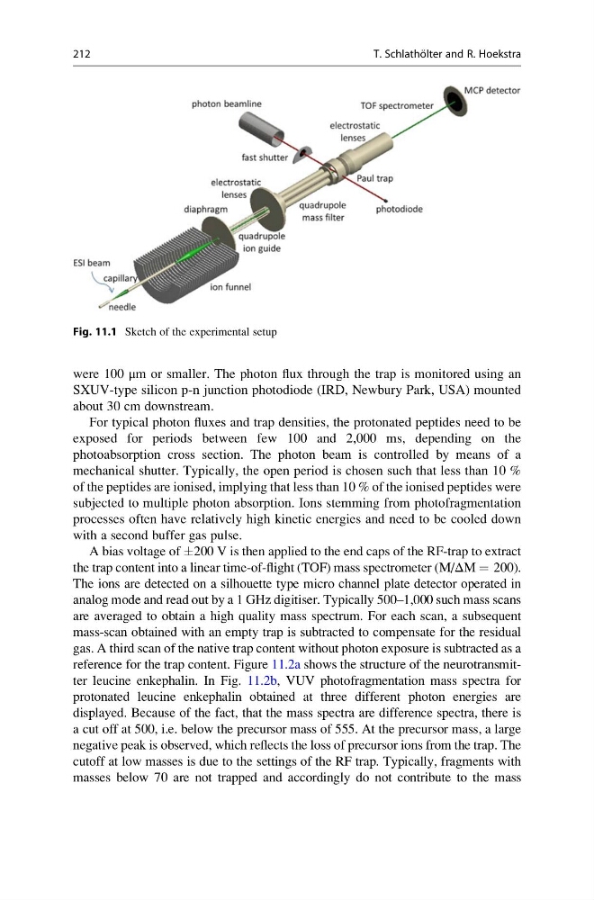
Fig. 11.1 Sketch of the experimental setup
212
T. Schlatho ¨ lter and R. Hoekstra
SXUV-type silicon p-n junction photodiode (IRD, Newbury Park, USA) mounted
about 30 cm downstream.
For typical photon fluxes and trap densities, the protonated peptides need to be
exposed for periods between few 100 and 2,000 ms, depending on the
photoabsorption cross section. The photon beam is controlled by means of a
mechanical shutter. Typically, the open period is chosen such that less than 10 %
of the peptides are ionised, implying that less than 10 % of the ionised peptides were
subjected to multiple photon absorption. Ions stemming from photofragmentation
processes often have relatively high kinetic energies and need to be cooled down
with a second buffer gas pulse.
A bias voltage of Æ200 V is then applied to the end caps of the RF-trap to extract
the trap content into a linear time-of-flight (TOF) mass spectrometer (M/ΔM ¼ 200).
The ions are detected on a silhouette type micro channel plate detector operated in
analog mode and read out by a 1 GHz digitiser. Typically 500–1,000 such mass scans
are averaged to obtain a high quality mass spectrum. For each scan, a subsequent
mass-scan obtained with an empty trap is subtracted to compensate for the residual
gas. A third scan of the native trap content without photon exposure is subtracted as a
reference for the trap content. Figure 11.2a shows the structure of the neurotransmitter leucine enkephalin. In Fig. 11.2b, VUV photofragmentation mass spectra for
protonated leucine enkephalin obtained at three different photon energies are
displayed. Because of the fact, that the mass spectra are difference spectra, there is
a cut off at 500, i.e. below the precursor mass of 555. At the precursor mass, a large
negative peak is observed, which reflects the loss of precursor ions from the trap. The
cutoff at low masses is due to the settings of the RF trap. Typically, fragments with
masses below 70 are not trapped and accordingly do not contribute to the mass
Fig. 11.1 Sketch of the experimental setup
212
T. Schlatho ¨ lter and R. Hoekstra