

formation of B
À was found to be the dominant fragmentation process for the cyclic
mononucleotides. This presents several questions as to the mechanisms that are
involved. If the previously proposed E2-type mechanism (Scheme 10.3) is still at
play, one of the two phosphoric acid ester bridges must be broken first to allow
glycosidic bond cleavage and concomitant proton transfer. In addition, the observed
formation of PO 3
À and H 2 PO 4
À fragments means that at least two bonds between
the phosphate and sugar groups must be cleaved. If one of the phosphate ester
bridges is first broken so that the phosphate group can participate in an E2-type
reaction, the preference of B
À loss over BH could be explained by the smaller
number of acidic protons available for transfer to the nucleobase in cyclic
nucleotides. Alternative mechanisms are discussed in more detail in [27, 28], but
no single reaction scheme can consistently explain all observations, similar to the
complex processes encountered in CID [43].
10.3 Time Scales for Dissociation
An interesting question is whether or not photoproduct formation, as described in
the preceding section, can compete with relaxation by energy transfer to solvent
when the nucleotide is solvated. If prompt dissociation processes are possible, they
would be hazardous to biological systems. However, if dissociation occurs on long
time scales, the more rapid energy transfer to solvent (typically within a few
picoseconds [1]) will most likely preclude significant amounts of fragmentation.
Thus, to determine whether or not fragmentation is important in biological systems,
it is necessary to switch off energy dissipation to the solvent and elucidate the
intrinsic dissociation times of nucleotides so that they can be compared to time
scales for other relaxation mechanisms. Measurement of such intrinsic dissociation
timescales can be accomplished in vacuo using mass spectrometric experiments.
We note that the measurement of decay times on the scale of 10
À8 to 10
À3 s is
difficult, since they are too long for typical optical delay line techniques and too
short to employ ion trapping approaches, e.g., in Paul traps or Fourier-transform
ion-cyclotron resonance mass spectrometers. Such experiments are best performed
using ion optical approaches and ion storage devices (see, e.g., [47–50]).
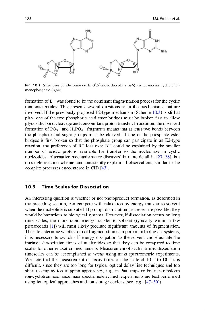
Fig. 10.2 Structures of adenosine cyclic-3
0 ,5
0 -monophosphate (left) and guanosine cyclic-3
0 ,5
0 -
monophosphate (right)
188
J.M. Weber et al.
À was found to be the dominant fragmentation process for the cyclic
mononucleotides. This presents several questions as to the mechanisms that are
involved. If the previously proposed E2-type mechanism (Scheme 10.3) is still at
play, one of the two phosphoric acid ester bridges must be broken first to allow
glycosidic bond cleavage and concomitant proton transfer. In addition, the observed
formation of PO 3
À and H 2 PO 4
À fragments means that at least two bonds between
the phosphate and sugar groups must be cleaved. If one of the phosphate ester
bridges is first broken so that the phosphate group can participate in an E2-type
reaction, the preference of B
À loss over BH could be explained by the smaller
number of acidic protons available for transfer to the nucleobase in cyclic
nucleotides. Alternative mechanisms are discussed in more detail in [27, 28], but
no single reaction scheme can consistently explain all observations, similar to the
complex processes encountered in CID [43].
10.3 Time Scales for Dissociation
An interesting question is whether or not photoproduct formation, as described in
the preceding section, can compete with relaxation by energy transfer to solvent
when the nucleotide is solvated. If prompt dissociation processes are possible, they
would be hazardous to biological systems. However, if dissociation occurs on long
time scales, the more rapid energy transfer to solvent (typically within a few
picoseconds [1]) will most likely preclude significant amounts of fragmentation.
Thus, to determine whether or not fragmentation is important in biological systems,
it is necessary to switch off energy dissipation to the solvent and elucidate the
intrinsic dissociation times of nucleotides so that they can be compared to time
scales for other relaxation mechanisms. Measurement of such intrinsic dissociation
timescales can be accomplished in vacuo using mass spectrometric experiments.
We note that the measurement of decay times on the scale of 10
À8 to 10
À3 s is
difficult, since they are too long for typical optical delay line techniques and too
short to employ ion trapping approaches, e.g., in Paul traps or Fourier-transform
ion-cyclotron resonance mass spectrometers. Such experiments are best performed
using ion optical approaches and ion storage devices (see, e.g., [47–50]).
Fig. 10.2 Structures of adenosine cyclic-3
0 ,5
0 -monophosphate (left) and guanosine cyclic-3
0 ,5
0 -
monophosphate (right)
188
J.M. Weber et al.