

refers to the different folding motifs of the substructures, e.g., α-helix, β-sheet, and
random coil. How the substructures are organised relative to each other in three
dimensions is called the tertiary structure. If a protein has more than one polypeptide chain, then the quaternary structure is the arrangement of the polypeptides
relative to each other.
In gas phase, peptide basic groups, e.g. amino and guanidine groups, are easily
protonated forming positively charged cations, while negatively charged anions are
formed by deprotonation of carboxylic acid groups. In mass spectrometry the
sequence of a peptide is determined by fragmentation, induced by either collisions
after acceleration, electron capture, or photoexcitation, followed by mass spectrometric analysis of the fragment ions. The nomenclature for fragment ions is
illustrated for a tetrapeptide in Fig. 1.10.
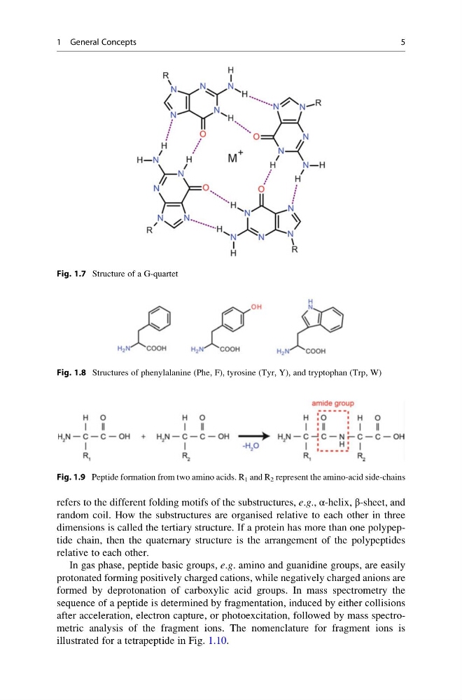
Fig. 1.7 Structure of a G-quartet
Fig. 1.8 Structures of phenylalanine (Phe, F), tyrosine (Tyr, Y), and tryptophan (Trp, W)
Fig. 1.9 Peptide formation from two amino acids. R 1 and R 2 represent the amino-acid side-chains
1 General Concepts
5
random coil. How the substructures are organised relative to each other in three
dimensions is called the tertiary structure. If a protein has more than one polypeptide chain, then the quaternary structure is the arrangement of the polypeptides
relative to each other.
In gas phase, peptide basic groups, e.g. amino and guanidine groups, are easily
protonated forming positively charged cations, while negatively charged anions are
formed by deprotonation of carboxylic acid groups. In mass spectrometry the
sequence of a peptide is determined by fragmentation, induced by either collisions
after acceleration, electron capture, or photoexcitation, followed by mass spectrometric analysis of the fragment ions. The nomenclature for fragment ions is
illustrated for a tetrapeptide in Fig. 1.10.
Fig. 1.7 Structure of a G-quartet
Fig. 1.8 Structures of phenylalanine (Phe, F), tyrosine (Tyr, Y), and tryptophan (Trp, W)
Fig. 1.9 Peptide formation from two amino acids. R 1 and R 2 represent the amino-acid side-chains
1 General Concepts
5