

abundance. These fragments are secondary fragments and are obtained in CID
when the collision energy is increased.
The change in fragmentation channels, not all observed in CID, shows that the
initially excited state has a strong influence on the fragmentation dynamics, and that
the dynamics is more complex than internal conversion followed by statistical
dissociation. More information is needed to explain these fragmentations.
At higher spectral resolution, one can see that conformer A of Tyr (the
conformer with the lowest transition energy) does not have the same fragmentation
pattern as isomer B. This is reflected in Fig. 9.7 in which the observed spectrum
depends on the fragment detected. This is a very important observation, which
shows that the fragmentation process is not at all statistical: the energy brought by
the laser for all the isomers is the same within 0.1 % since the electronic transitions
are very similar and isomer B (and D) are leading more efficiently to the m/z ¼ 107
or 108 fragment. As seen in the experiment of Fig. 9.6, the fragment is m/z ¼ 108
which corresponds to the side-chain bond rupture after a proton transfer from the
ammonium group towards the phenol ring (a simple C α –C β bond rupture would lead
to m/z ¼ 107). The fragmentation is driven by the initial structure, which also
implies that the electronic properties and the mechanisms are strongly dependent on
small changes of geometry. The structures of these isomers were identified from IR/
UV [18] double resonance spectra in combination with ab-initio ground state
calculations (see Fig. 9.8). In isomer A and C, both the NH 3
+ and CO groups are
above the phenol ring whereas in isomer B and D (B is the most stable isomer by
only 2.7 kJ/mol) only the ammonium group is above the ring. A and C are quite
similar with respect to the rotation around the C α –C β bond, they differ only by the
flipping of the OH bend of the phenol moiety and it is similar for isomers B and D.
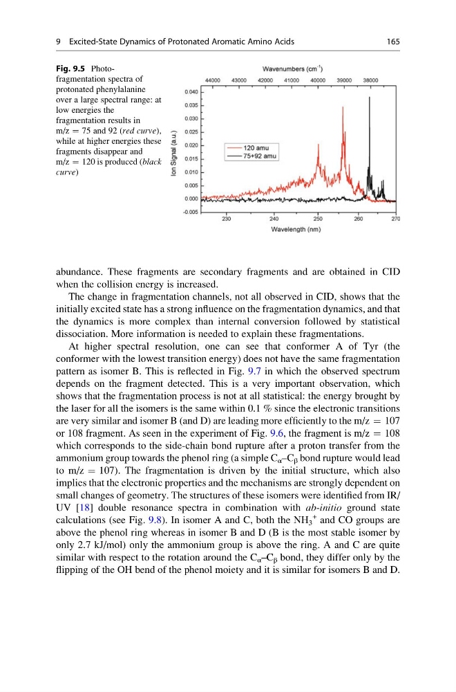
Fig. 9.5 Photofragmentation spectra of
protonated phenylalanine
over a large spectral range: at
low energies the
fragmentation results in
m/z ¼ 75 and 92 (red curve),
while at higher energies these
fragments disappear and
m/z ¼ 120 is produced (black
curve)
9 Excited-State Dynamics of Protonated Aromatic Amino Acids
165
when the collision energy is increased.
The change in fragmentation channels, not all observed in CID, shows that the
initially excited state has a strong influence on the fragmentation dynamics, and that
the dynamics is more complex than internal conversion followed by statistical
dissociation. More information is needed to explain these fragmentations.
At higher spectral resolution, one can see that conformer A of Tyr (the
conformer with the lowest transition energy) does not have the same fragmentation
pattern as isomer B. This is reflected in Fig. 9.7 in which the observed spectrum
depends on the fragment detected. This is a very important observation, which
shows that the fragmentation process is not at all statistical: the energy brought by
the laser for all the isomers is the same within 0.1 % since the electronic transitions
are very similar and isomer B (and D) are leading more efficiently to the m/z ¼ 107
or 108 fragment. As seen in the experiment of Fig. 9.6, the fragment is m/z ¼ 108
which corresponds to the side-chain bond rupture after a proton transfer from the
ammonium group towards the phenol ring (a simple C α –C β bond rupture would lead
to m/z ¼ 107). The fragmentation is driven by the initial structure, which also
implies that the electronic properties and the mechanisms are strongly dependent on
small changes of geometry. The structures of these isomers were identified from IR/
UV [18] double resonance spectra in combination with ab-initio ground state
calculations (see Fig. 9.8). In isomer A and C, both the NH 3
+ and CO groups are
above the phenol ring whereas in isomer B and D (B is the most stable isomer by
only 2.7 kJ/mol) only the ammonium group is above the ring. A and C are quite
similar with respect to the rotation around the C α –C β bond, they differ only by the
flipping of the OH bend of the phenol moiety and it is similar for isomers B and D.
Fig. 9.5 Photofragmentation spectra of
protonated phenylalanine
over a large spectral range: at
low energies the
fragmentation results in
m/z ¼ 75 and 92 (red curve),
while at higher energies these
fragments disappear and
m/z ¼ 120 is produced (black
curve)
9 Excited-State Dynamics of Protonated Aromatic Amino Acids
165