

8.3
Photofragmentation and Optical Spectroscopy of Trapped
Polypeptide and Protein Ions
Due to very low ion concentrations, absorption [13] and fluorescence [14]
spectroscopies of trapped ions have been reduced to a few examples, and gasphase spectroscopy most often relies on action spectra [11]. The principle is to use
the dissociation that follows photon absorption to monitor the optical properties of
the selected ion. A common assumption is that the photodissociation yield is
proportional to the absorption cross section.
The quest to perform spectroscopy on large systems in the gas phase, required to
ensure the biological relevance of gas-phase studies, was slowed down by the
bottleneck of action spectroscopy. As the size of the system and then the number
of degrees of freedom increases, the redistribution of vibrational energy that
follows internal conversion leads to a very small increase in temperature without
fragmentation in the time window of the experiment. The possibility of fragmentation from electronic excited states prior to internal vibrational energy redistribution
(IVR) [15] (see Fig. 8.2) may lead to the measurement of optical spectra for large
systems. A possible drawback of this method may be the strong dependence of the
fragmentation rate on the structure of the ions and/or on the initial electronic
excited states, leading to a poor match between fragmentation yields and absorption
cross sections.
An alternative which was developed in Lyon is to work with polyanions. The
mass spectrum obtained after UV-laser irradiation at 260 nm of the doubly
deprotonated [M-2H]
2À gramicidin anion is shown in Fig. 8.3a. The main daughter
ion corresponds to the oxidised [M–2H]
À• ion generated by electron detachment
from the [M–2H]
2À precursor ion. This electron loss is a typical de-excitation
pathway for polyanions [16, 17]. Figure 8.3b presents the electron photodetachment
efficiency as a function of wavelength for the gramicidin peptide dianions. The
detachment can be seen below 300 nm where the absorption occurs in solution
(inset in Fig. 8.3b) due to ππ* excitations of aromatic residues. Thus, the first step
in this electron loss was a resonant electronic excitation of the precursor ion.
Absorption of a 260-nm photon by the peptide corresponds to an increase in energy
Ion source
Vacuum manifold
Ion trap
UV/Visible
Laser
OpƟcal window
Ion transfer
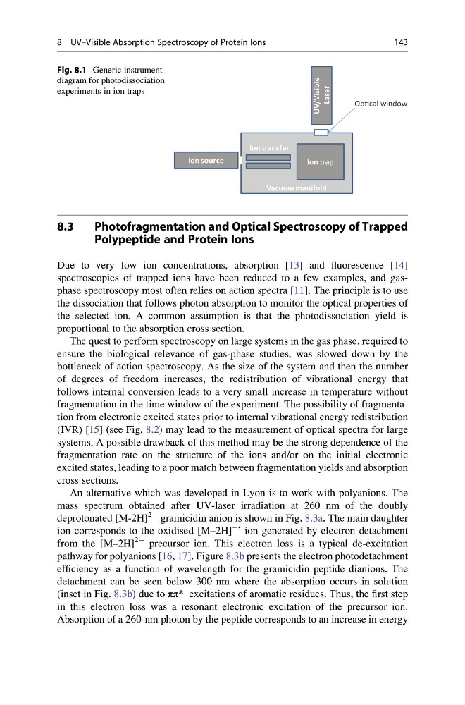
Fig. 8.1 Generic instrument
diagram for photodissociation
experiments in ion traps
8 UV–Visible Absorption Spectroscopy of Protein Ions
143
Photofragmentation and Optical Spectroscopy of Trapped
Polypeptide and Protein Ions
Due to very low ion concentrations, absorption [13] and fluorescence [14]
spectroscopies of trapped ions have been reduced to a few examples, and gasphase spectroscopy most often relies on action spectra [11]. The principle is to use
the dissociation that follows photon absorption to monitor the optical properties of
the selected ion. A common assumption is that the photodissociation yield is
proportional to the absorption cross section.
The quest to perform spectroscopy on large systems in the gas phase, required to
ensure the biological relevance of gas-phase studies, was slowed down by the
bottleneck of action spectroscopy. As the size of the system and then the number
of degrees of freedom increases, the redistribution of vibrational energy that
follows internal conversion leads to a very small increase in temperature without
fragmentation in the time window of the experiment. The possibility of fragmentation from electronic excited states prior to internal vibrational energy redistribution
(IVR) [15] (see Fig. 8.2) may lead to the measurement of optical spectra for large
systems. A possible drawback of this method may be the strong dependence of the
fragmentation rate on the structure of the ions and/or on the initial electronic
excited states, leading to a poor match between fragmentation yields and absorption
cross sections.
An alternative which was developed in Lyon is to work with polyanions. The
mass spectrum obtained after UV-laser irradiation at 260 nm of the doubly
deprotonated [M-2H]
2À gramicidin anion is shown in Fig. 8.3a. The main daughter
ion corresponds to the oxidised [M–2H]
À• ion generated by electron detachment
from the [M–2H]
2À precursor ion. This electron loss is a typical de-excitation
pathway for polyanions [16, 17]. Figure 8.3b presents the electron photodetachment
efficiency as a function of wavelength for the gramicidin peptide dianions. The
detachment can be seen below 300 nm where the absorption occurs in solution
(inset in Fig. 8.3b) due to ππ* excitations of aromatic residues. Thus, the first step
in this electron loss was a resonant electronic excitation of the precursor ion.
Absorption of a 260-nm photon by the peptide corresponds to an increase in energy
Ion source
Vacuum manifold
Ion trap
UV/Visible
Laser
OpƟcal window
Ion transfer
Fig. 8.1 Generic instrument
diagram for photodissociation
experiments in ion traps
8 UV–Visible Absorption Spectroscopy of Protein Ions
143