

In other experiments, 535-nm light was used as the pump, and the probe laser
wavelength was scanned. Furthermore, results for two different time delays between
the laser pulses were obtained (Fig. 7.19). Action spectra were then compiled by
analysing the increased yield of photofragments induced by the second laser pulse.
From both thus obtained spectra, it is evident that absorption is strong at 650 nm, and
that it decreases as the wavelength increases, with little or no absorption seen above
800 nm. Moreover, the band appears sharper after the longer time delay (0.57 ms)
than after the shorter one (0.19 ms), which may be due to a colder population at
longer delay times. Experiments at lower probe wavelengths were complicated by
ground-state absorption (see Fig. 7.19a), which means that it is difficult to exclude
that there is some contribution from the Q 0 band, estimated to be maximal at 630 nm,
to the pump-probe absorption spectrum at the lowest wavelengths.
5
10
15
20
Time (ms)
Counts / 100
35
36
37
38
5
10
15
20
10
20
30
40
c
b
a
t 2 = 0.87 ms
t 1 = 67 ms
Pump
Probe
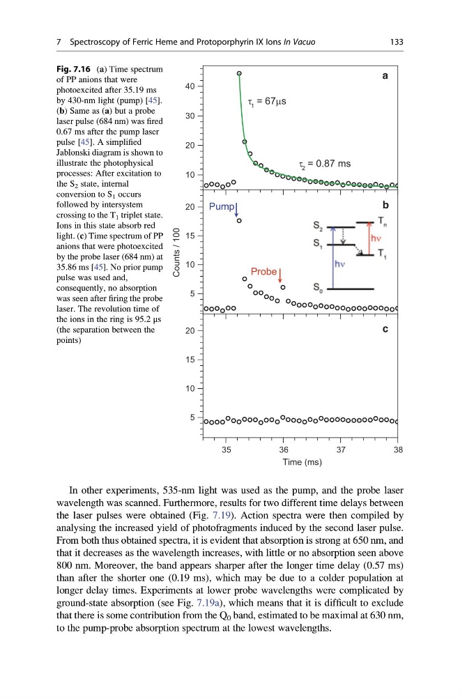
Fig. 7.16 (a) Time spectrum
of PP anions that were
photoexcited after 35.19 ms
by 430-nm light (pump) [45].
(b) Same as (a) but a probe
laser pulse (684 nm) was fired
0.67 ms after the pump laser
pulse [45]. A simplified
Jablonski diagram is shown to
illustrate the photophysical
processes: After excitation to
the S 2 state, internal
conversion to S 1 occurs
followed by intersystem
crossing to the T 1 triplet state.
Ions in this state absorb red
light. (c) Time spectrum of PP
anions that were photoexcited
by the probe laser (684 nm) at
35.86 ms [45]. No prior pump
pulse was used and,
consequently, no absorption
was seen after firing the probe
laser. The revolution time of
the ions in the ring is 95.2 μs
(the separation between the
points)
7 Spectroscopy of Ferric Heme and Protoporphyrin IX Ions In Vacuo
133
wavelength was scanned. Furthermore, results for two different time delays between
the laser pulses were obtained (Fig. 7.19). Action spectra were then compiled by
analysing the increased yield of photofragments induced by the second laser pulse.
From both thus obtained spectra, it is evident that absorption is strong at 650 nm, and
that it decreases as the wavelength increases, with little or no absorption seen above
800 nm. Moreover, the band appears sharper after the longer time delay (0.57 ms)
than after the shorter one (0.19 ms), which may be due to a colder population at
longer delay times. Experiments at lower probe wavelengths were complicated by
ground-state absorption (see Fig. 7.19a), which means that it is difficult to exclude
that there is some contribution from the Q 0 band, estimated to be maximal at 630 nm,
to the pump-probe absorption spectrum at the lowest wavelengths.
5
10
15
20
Time (ms)
Counts / 100
35
36
37
38
5
10
15
20
10
20
30
40
c
b
a
t 2 = 0.87 ms
t 1 = 67 ms
Pump
Probe
Fig. 7.16 (a) Time spectrum
of PP anions that were
photoexcited after 35.19 ms
by 430-nm light (pump) [45].
(b) Same as (a) but a probe
laser pulse (684 nm) was fired
0.67 ms after the pump laser
pulse [45]. A simplified
Jablonski diagram is shown to
illustrate the photophysical
processes: After excitation to
the S 2 state, internal
conversion to S 1 occurs
followed by intersystem
crossing to the T 1 triplet state.
Ions in this state absorb red
light. (c) Time spectrum of PP
anions that were photoexcited
by the probe laser (684 nm) at
35.86 ms [45]. No prior pump
pulse was used and,
consequently, no absorption
was seen after firing the probe
laser. The revolution time of
the ions in the ring is 95.2 μs
(the separation between the
points)
7 Spectroscopy of Ferric Heme and Protoporphyrin IX Ions In Vacuo
133