

7.3
Time Constants and Channels for Photo-induced
Dissociation
Dissociation of photoexcited Fe(III)–heme
+ and Fe(III)–heme
+ (His) ions occurs on
the microsecond to millisecond time scale with the main channels being ascribed to
loss of either one or two CH 2 COOH groups through β-cleavage, and loss of
histidine, respectively (see Figs. 7.13 and 7.14) [31]. The former reaction is
associated with a reaction energy of about 2 eV while histidine loss costs less
energy, only about 1.4 eV. In agreement with this, the dissociation of Fe(III)–heme
+
was found to result from the absorption of either one or two photons, somewhat
dependent on the excitation wavelength, while that of Fe(III)–heme
+ (His) could be
accounted for purely by one-photon absorption. In this regard, the formation of the
m/z-498 fragment ion from heme
+ (loss of two CH 2 COOH groups) right after
photoexcitation is clear evidence of two-photon absorption as one visible photon
is not enough to cause loss of both CH 2 COOH groups (Fig. 7.14c).
0
1
2
3
4
5
0
1
2
3
4
5
Absorption (arb. units)
b
a
Protein absorption
450
500
550
600
650
700
Wavelength (nm)
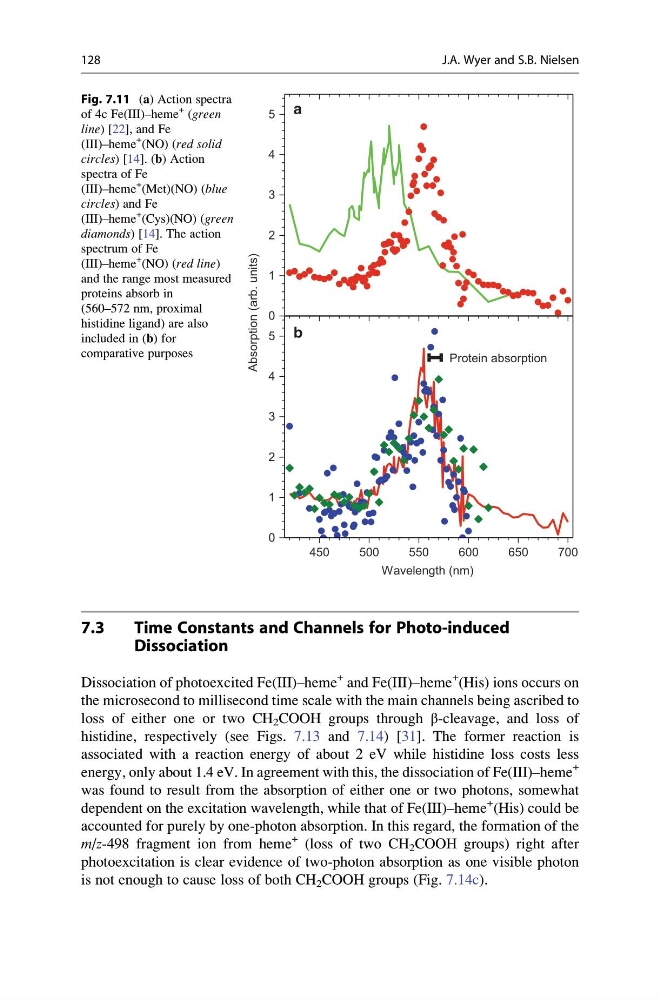
Fig. 7.11 (a) Action spectra
of 4c Fe(III)–heme
+ (green
line) [22], and Fe
(III)–heme
+ (NO) (red solid
circles) [14]. (b) Action
spectra of Fe
(III)–heme
+ (Met)(NO) (blue
circles) and Fe
(III)–heme
+ (Cys)(NO) (green
diamonds) [14]. The action
spectrum of Fe
(III)–heme
+ (NO) (red line)
and the range most measured
proteins absorb in
(560–572 nm, proximal
histidine ligand) are also
included in (b) for
comparative purposes
128
J.A. Wyer and S.B. Nielsen
Time Constants and Channels for Photo-induced
Dissociation
Dissociation of photoexcited Fe(III)–heme
+ and Fe(III)–heme
+ (His) ions occurs on
the microsecond to millisecond time scale with the main channels being ascribed to
loss of either one or two CH 2 COOH groups through β-cleavage, and loss of
histidine, respectively (see Figs. 7.13 and 7.14) [31]. The former reaction is
associated with a reaction energy of about 2 eV while histidine loss costs less
energy, only about 1.4 eV. In agreement with this, the dissociation of Fe(III)–heme
+
was found to result from the absorption of either one or two photons, somewhat
dependent on the excitation wavelength, while that of Fe(III)–heme
+ (His) could be
accounted for purely by one-photon absorption. In this regard, the formation of the
m/z-498 fragment ion from heme
+ (loss of two CH 2 COOH groups) right after
photoexcitation is clear evidence of two-photon absorption as one visible photon
is not enough to cause loss of both CH 2 COOH groups (Fig. 7.14c).
0
1
2
3
4
5
0
1
2
3
4
5
Absorption (arb. units)
b
a
Protein absorption
450
500
550
600
650
700
Wavelength (nm)
Fig. 7.11 (a) Action spectra
of 4c Fe(III)–heme
+ (green
line) [22], and Fe
(III)–heme
+ (NO) (red solid
circles) [14]. (b) Action
spectra of Fe
(III)–heme
+ (Met)(NO) (blue
circles) and Fe
(III)–heme
+ (Cys)(NO) (green
diamonds) [14]. The action
spectrum of Fe
(III)–heme
+ (NO) (red line)
and the range most measured
proteins absorb in
(560–572 nm, proximal
histidine ligand) are also
included in (b) for
comparative purposes
128
J.A. Wyer and S.B. Nielsen