

Fig. 7.7 dd excited states act as doorway states, quickly bringing the heme back to its electronic
ground state. Note vibrational levels are not included for clarity
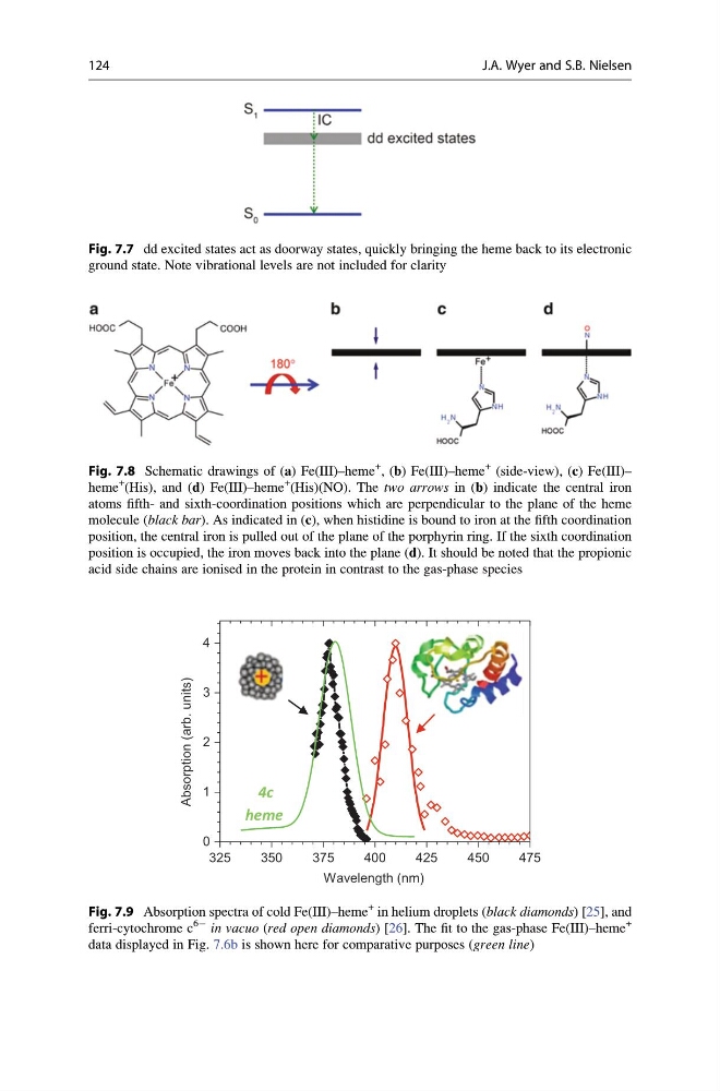
Fig. 7.8 Schematic drawings of (a) Fe(III)–heme
+ , (b) Fe(III)–heme
+ (side-view), (c) Fe(III)–
heme
+ (His), and (d) Fe(III)–heme
+ (His)(NO). The two arrows in (b) indicate the central iron
atoms fifth- and sixth-coordination positions which are perpendicular to the plane of the heme
molecule (black bar). As indicated in (c), when histidine is bound to iron at the fifth coordination
position, the central iron is pulled out of the plane of the porphyrin ring. If the sixth coordination
position is occupied, the iron moves back into the plane (d). It should be noted that the propionic
acid side chains are ionised in the protein in contrast to the gas-phase species
325
350
375
400
425
450
475
0
1
2
3
4
Absorption (arb. units)
Wavelength (nm)
4c
heme
Fig. 7.9 Absorption spectra of cold Fe(III)–heme
+ in helium droplets (black diamonds) [25], and
ferri-cytochrome c
6À in vacuo (red open diamonds) [26]. The fit to the gas-phase Fe(III)–heme
+
data displayed in Fig. 7.6b is shown here for comparative purposes (green line)
124
J.A. Wyer and S.B. Nielsen
ground state. Note vibrational levels are not included for clarity
Fig. 7.8 Schematic drawings of (a) Fe(III)–heme
+ , (b) Fe(III)–heme
+ (side-view), (c) Fe(III)–
heme
+ (His), and (d) Fe(III)–heme
+ (His)(NO). The two arrows in (b) indicate the central iron
atoms fifth- and sixth-coordination positions which are perpendicular to the plane of the heme
molecule (black bar). As indicated in (c), when histidine is bound to iron at the fifth coordination
position, the central iron is pulled out of the plane of the porphyrin ring. If the sixth coordination
position is occupied, the iron moves back into the plane (d). It should be noted that the propionic
acid side chains are ionised in the protein in contrast to the gas-phase species
325
350
375
400
425
450
475
0
1
2
3
4
Absorption (arb. units)
Wavelength (nm)
4c
heme
Fig. 7.9 Absorption spectra of cold Fe(III)–heme
+ in helium droplets (black diamonds) [25], and
ferri-cytochrome c
6À in vacuo (red open diamonds) [26]. The fit to the gas-phase Fe(III)–heme
+
data displayed in Fig. 7.6b is shown here for comparative purposes (green line)
124
J.A. Wyer and S.B. Nielsen