

ring, pulling the histidine, which causes a strain that induces conformational
changes in the protein. O 2 and CO only bind to ferrous heme, while the NO radical
can coordinate to both ferrous and ferric heme. These diatomic molecules activate
or inhibit key biological processes by binding to heme, but the processes strongly
depend on the surrounding protein environment that modulates the electronic
properties of the adduct. Both electrostatic interactions and hydrogen-bonding
effects associated with sidechains distal to the bound molecule can play a role.
Furthermore, the axial ligand trans to the diatomic molecule is important, often
being either histidine (cf., Mb and Hb) or cysteine. The strength of its binding
influences the bond between iron and the diatomic molecule.
Heme–NO proteins play a key role in many physiological functions including
blood clotting, blood pressure regulation, immune response, nerve signal transduction, and vasodilation upon the bite of blood-sucking insects [6–8]. Even though
ferric heme can bind NO, the affinity for NO is lower for Fe(III) than Fe(II), a fact
that is exploited in the liberation of NO after transportation [9]. Furthermore, the Fe
(II)–NO and Fe(III)–NO moieties are different, with the former adopting a bent
conformation and the latter a linear one when the proximal ligand is histidine
[9–12]. As this linearity is caused by electron transfer from NO to Fe(III) through
the σ orbital a more proper description of the unit is Fe(II)–NO
+ [9–12]. According
to TD-DFT and CASSCF calculations this is a singlet state [11, 13]. The absorption
in the Q band region in measured ferric nitrosyl proteins ranges from 549 nm to
574 nm in the Q 0 band and 518 nm to 538 nm in the Q 1 band (see Table in [14]). If,
on the other hand, the proximal ligand is an electron-rich ligand such as cysteinate,
then the electron donation is retarded and back-donation from the iron to NO is
facilitated. This results in a slightly bent and tilted coordination [10, 11], and the
absorption in the Q-band region is redshifted to 571–585 nm (Q 0 ) and 533–549 nm
(Q 1 ). For both ferric heme nitrosyl complexes, the Enemark-Feltham notation is
{Fe(NO)}
6 where 6 refers to the number of Fe d electrons plus the unpaired electron
of NO.
200
300
400
500
600
700
800
0.0
0.2
0.4
0.6
0.8
1.0
Wavelength (nm)
Absorption (arb. units)
Q 0
Soret
band
Q 1
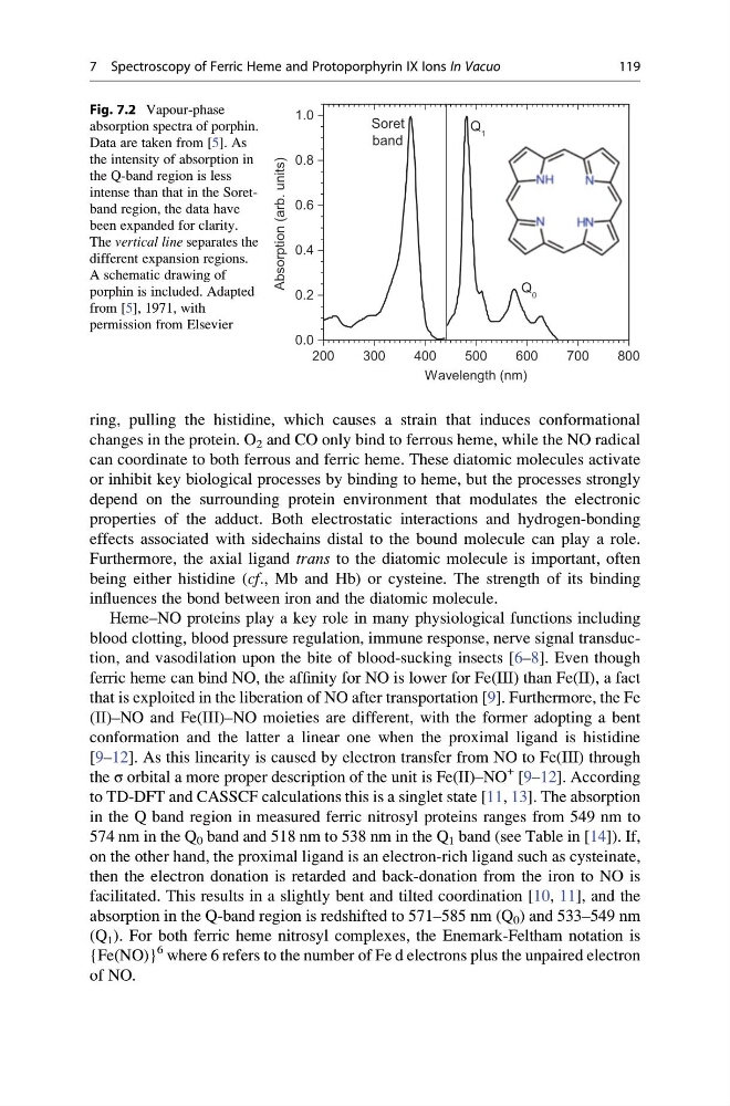
Fig. 7.2 Vapour-phase
absorption spectra of porphin.
Data are taken from [5]. As
the intensity of absorption in
the Q-band region is less
intense than that in the Soretband region, the data have
been expanded for clarity.
The vertical line separates the
different expansion regions.
A schematic drawing of
porphin is included. Adapted
from [5], 1971, with
permission from Elsevier
7 Spectroscopy of Ferric Heme and Protoporphyrin IX Ions In Vacuo
119
changes in the protein. O 2 and CO only bind to ferrous heme, while the NO radical
can coordinate to both ferrous and ferric heme. These diatomic molecules activate
or inhibit key biological processes by binding to heme, but the processes strongly
depend on the surrounding protein environment that modulates the electronic
properties of the adduct. Both electrostatic interactions and hydrogen-bonding
effects associated with sidechains distal to the bound molecule can play a role.
Furthermore, the axial ligand trans to the diatomic molecule is important, often
being either histidine (cf., Mb and Hb) or cysteine. The strength of its binding
influences the bond between iron and the diatomic molecule.
Heme–NO proteins play a key role in many physiological functions including
blood clotting, blood pressure regulation, immune response, nerve signal transduction, and vasodilation upon the bite of blood-sucking insects [6–8]. Even though
ferric heme can bind NO, the affinity for NO is lower for Fe(III) than Fe(II), a fact
that is exploited in the liberation of NO after transportation [9]. Furthermore, the Fe
(II)–NO and Fe(III)–NO moieties are different, with the former adopting a bent
conformation and the latter a linear one when the proximal ligand is histidine
[9–12]. As this linearity is caused by electron transfer from NO to Fe(III) through
the σ orbital a more proper description of the unit is Fe(II)–NO
+ [9–12]. According
to TD-DFT and CASSCF calculations this is a singlet state [11, 13]. The absorption
in the Q band region in measured ferric nitrosyl proteins ranges from 549 nm to
574 nm in the Q 0 band and 518 nm to 538 nm in the Q 1 band (see Table in [14]). If,
on the other hand, the proximal ligand is an electron-rich ligand such as cysteinate,
then the electron donation is retarded and back-donation from the iron to NO is
facilitated. This results in a slightly bent and tilted coordination [10, 11], and the
absorption in the Q-band region is redshifted to 571–585 nm (Q 0 ) and 533–549 nm
(Q 1 ). For both ferric heme nitrosyl complexes, the Enemark-Feltham notation is
{Fe(NO)}
6 where 6 refers to the number of Fe d electrons plus the unpaired electron
of NO.
200
300
400
500
600
700
800
0.0
0.2
0.4
0.6
0.8
1.0
Wavelength (nm)
Absorption (arb. units)
Q 0
Soret
band
Q 1
Fig. 7.2 Vapour-phase
absorption spectra of porphin.
Data are taken from [5]. As
the intensity of absorption in
the Q-band region is less
intense than that in the Soretband region, the data have
been expanded for clarity.
The vertical line separates the
different expansion regions.
A schematic drawing of
porphin is included. Adapted
from [5], 1971, with
permission from Elsevier
7 Spectroscopy of Ferric Heme and Protoporphyrin IX Ions In Vacuo
119