

7.1
Introduction
Porphyrin-molecules are ubiquitous in nature and are engaged in many essential
biological processes, e.g., photosynthesis, dioxygen transport and storage, signalling and sensing [1–3]. They belong to a class of highly symmetric heterocyclic
π-conjugated systems composed of the parent unit porphin with various peripheral
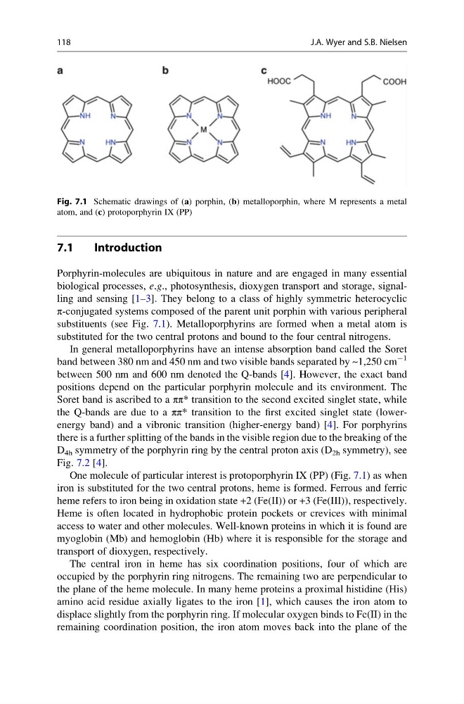
substituents (see Fig. 7.1). Metalloporphyrins are formed when a metal atom is
substituted for the two central protons and bound to the four central nitrogens.
In general metalloporphyrins have an intense absorption band called the Soret
band between 380 nm and 450 nm and two visible bands separated by ~1,250 cm
À1
between 500 nm and 600 nm denoted the Q-bands [4]. However, the exact band
positions depend on the particular porphyrin molecule and its environment. The
Soret band is ascribed to a ππ* transition to the second excited singlet state, while
the Q-bands are due to a ππ* transition to the first excited singlet state (lowerenergy band) and a vibronic transition (higher-energy band) [4]. For porphyrins
there is a further splitting of the bands in the visible region due to the breaking of the
D 4h symmetry of the porphyrin ring by the central proton axis (D 2h symmetry), see
Fig. 7.2 [4].
One molecule of particular interest is protoporphyrin IX (PP) (Fig. 7.1) as when
iron is substituted for the two central protons, heme is formed. Ferrous and ferric
heme refers to iron being in oxidation state +2 (Fe(II)) or +3 (Fe(III)), respectively.
Heme is often located in hydrophobic protein pockets or crevices with minimal
access to water and other molecules. Well-known proteins in which it is found are
myoglobin (Mb) and hemoglobin (Hb) where it is responsible for the storage and
transport of dioxygen, respectively.
The central iron in heme has six coordination positions, four of which are
occupied by the porphyrin ring nitrogens. The remaining two are perpendicular to
the plane of the heme molecule. In many heme proteins a proximal histidine (His)
amino acid residue axially ligates to the iron [1], which causes the iron atom to
displace slightly from the porphyrin ring. If molecular oxygen binds to Fe(II) in the
remaining coordination position, the iron atom moves back into the plane of the
Fig. 7.1 Schematic drawings of (a) porphin, (b) metalloporphin, where M represents a metal
atom, and (c) protoporphyrin IX (PP)
118
J.A. Wyer and S.B. Nielsen
Introduction
Porphyrin-molecules are ubiquitous in nature and are engaged in many essential
biological processes, e.g., photosynthesis, dioxygen transport and storage, signalling and sensing [1–3]. They belong to a class of highly symmetric heterocyclic
π-conjugated systems composed of the parent unit porphin with various peripheral
substituents (see Fig. 7.1). Metalloporphyrins are formed when a metal atom is
substituted for the two central protons and bound to the four central nitrogens.
In general metalloporphyrins have an intense absorption band called the Soret
band between 380 nm and 450 nm and two visible bands separated by ~1,250 cm
À1
between 500 nm and 600 nm denoted the Q-bands [4]. However, the exact band
positions depend on the particular porphyrin molecule and its environment. The
Soret band is ascribed to a ππ* transition to the second excited singlet state, while
the Q-bands are due to a ππ* transition to the first excited singlet state (lowerenergy band) and a vibronic transition (higher-energy band) [4]. For porphyrins
there is a further splitting of the bands in the visible region due to the breaking of the
D 4h symmetry of the porphyrin ring by the central proton axis (D 2h symmetry), see
Fig. 7.2 [4].
One molecule of particular interest is protoporphyrin IX (PP) (Fig. 7.1) as when
iron is substituted for the two central protons, heme is formed. Ferrous and ferric
heme refers to iron being in oxidation state +2 (Fe(II)) or +3 (Fe(III)), respectively.
Heme is often located in hydrophobic protein pockets or crevices with minimal
access to water and other molecules. Well-known proteins in which it is found are
myoglobin (Mb) and hemoglobin (Hb) where it is responsible for the storage and
transport of dioxygen, respectively.
The central iron in heme has six coordination positions, four of which are
occupied by the porphyrin ring nitrogens. The remaining two are perpendicular to
the plane of the heme molecule. In many heme proteins a proximal histidine (His)
amino acid residue axially ligates to the iron [1], which causes the iron atom to
displace slightly from the porphyrin ring. If molecular oxygen binds to Fe(II) in the
remaining coordination position, the iron atom moves back into the plane of the
Fig. 7.1 Schematic drawings of (a) porphin, (b) metalloporphin, where M represents a metal
atom, and (c) protoporphyrin IX (PP)
118
J.A. Wyer and S.B. Nielsen