

photophysical properties of the oxyluciferin anion from environmental
perturbations, work on isolated ions is needed.
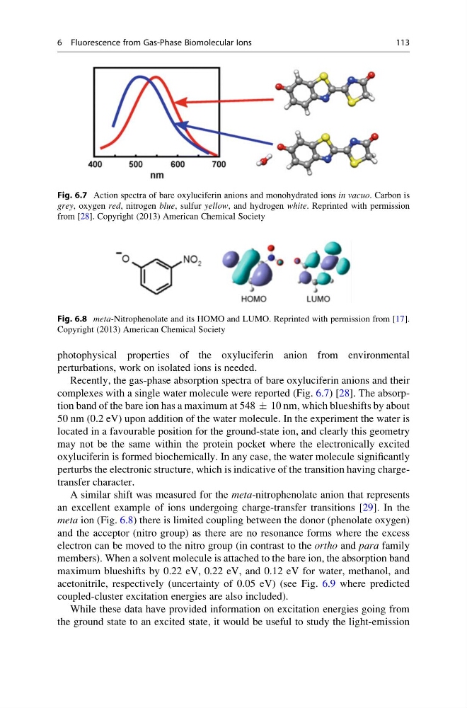
Recently, the gas-phase absorption spectra of bare oxyluciferin anions and their
complexes with a single water molecule were reported (Fig. 6.7) [28]. The absorption band of the bare ion has a maximum at 548 Æ 10 nm, which blueshifts by about
50 nm (0.2 eV) upon addition of the water molecule. In the experiment the water is
located in a favourable position for the ground-state ion, and clearly this geometry
may not be the same within the protein pocket where the electronically excited
oxyluciferin is formed biochemically. In any case, the water molecule significantly
perturbs the electronic structure, which is indicative of the transition having chargetransfer character.
A similar shift was measured for the meta-nitrophenolate anion that represents
an excellent example of ions undergoing charge-transfer transitions [29]. In the
meta ion (Fig. 6.8) there is limited coupling between the donor (phenolate oxygen)
and the acceptor (nitro group) as there are no resonance forms where the excess
electron can be moved to the nitro group (in contrast to the ortho and para family
members). When a solvent molecule is attached to the bare ion, the absorption band
maximum blueshifts by 0.22 eV, 0.22 eV, and 0.12 eV for water, methanol, and
acetonitrile, respectively (uncertainty of 0.05 eV) (see Fig. 6.9 where predicted
coupled-cluster excitation energies are also included).
While these data have provided information on excitation energies going from
the ground state to an excited state, it would be useful to study the light-emission
Fig. 6.7 Action spectra of bare oxyluciferin anions and monohydrated ions in vacuo. Carbon is
grey, oxygen red, nitrogen blue, sulfur yellow, and hydrogen white. Reprinted with permission
from [28]. Copyright (2013) American Chemical Society
Fig. 6.8 meta-Nitrophenolate and its HOMO and LUMO. Reprinted with permission from [17].
Copyright (2013) American Chemical Society
6 Fluorescence from Gas-Phase Biomolecular Ions
113
perturbations, work on isolated ions is needed.
Recently, the gas-phase absorption spectra of bare oxyluciferin anions and their
complexes with a single water molecule were reported (Fig. 6.7) [28]. The absorption band of the bare ion has a maximum at 548 Æ 10 nm, which blueshifts by about
50 nm (0.2 eV) upon addition of the water molecule. In the experiment the water is
located in a favourable position for the ground-state ion, and clearly this geometry
may not be the same within the protein pocket where the electronically excited
oxyluciferin is formed biochemically. In any case, the water molecule significantly
perturbs the electronic structure, which is indicative of the transition having chargetransfer character.
A similar shift was measured for the meta-nitrophenolate anion that represents
an excellent example of ions undergoing charge-transfer transitions [29]. In the
meta ion (Fig. 6.8) there is limited coupling between the donor (phenolate oxygen)
and the acceptor (nitro group) as there are no resonance forms where the excess
electron can be moved to the nitro group (in contrast to the ortho and para family
members). When a solvent molecule is attached to the bare ion, the absorption band
maximum blueshifts by 0.22 eV, 0.22 eV, and 0.12 eV for water, methanol, and
acetonitrile, respectively (uncertainty of 0.05 eV) (see Fig. 6.9 where predicted
coupled-cluster excitation energies are also included).
While these data have provided information on excitation energies going from
the ground state to an excited state, it would be useful to study the light-emission
Fig. 6.7 Action spectra of bare oxyluciferin anions and monohydrated ions in vacuo. Carbon is
grey, oxygen red, nitrogen blue, sulfur yellow, and hydrogen white. Reprinted with permission
from [28]. Copyright (2013) American Chemical Society
Fig. 6.8 meta-Nitrophenolate and its HOMO and LUMO. Reprinted with permission from [17].
Copyright (2013) American Chemical Society
6 Fluorescence from Gas-Phase Biomolecular Ions
113