

118
K. Kneipp et al.
2.4.2 Vibrational Spectroscopy of Protein Monolayers Using
SEIRA
Compared to SERS and particularly also compared to non-linear Raman methods,
the electromagnetic contribution for SEIRA signals is “modest” since the effect is
only proportional to the square of the field enhancement (see Fig. 2.3). However,
IR absorption and Raman scattering follow different symmetry selection rules and
can therefore probe different vibrational modes. For a comprehensive vibrational
characterization, Raman scattering and IR absorption spectra are of interest.
As we have discussed, infrared absorption can be efficiently supported by nanoantenna arrays. The collective resonant excitation of the nanoantenna ensemble results
in signal enhancement factors of 10 4 − 10 5 . This “collectively enhanced IR absorption” (CEIRA) spectroscopy technique allows direct identification of vibrational
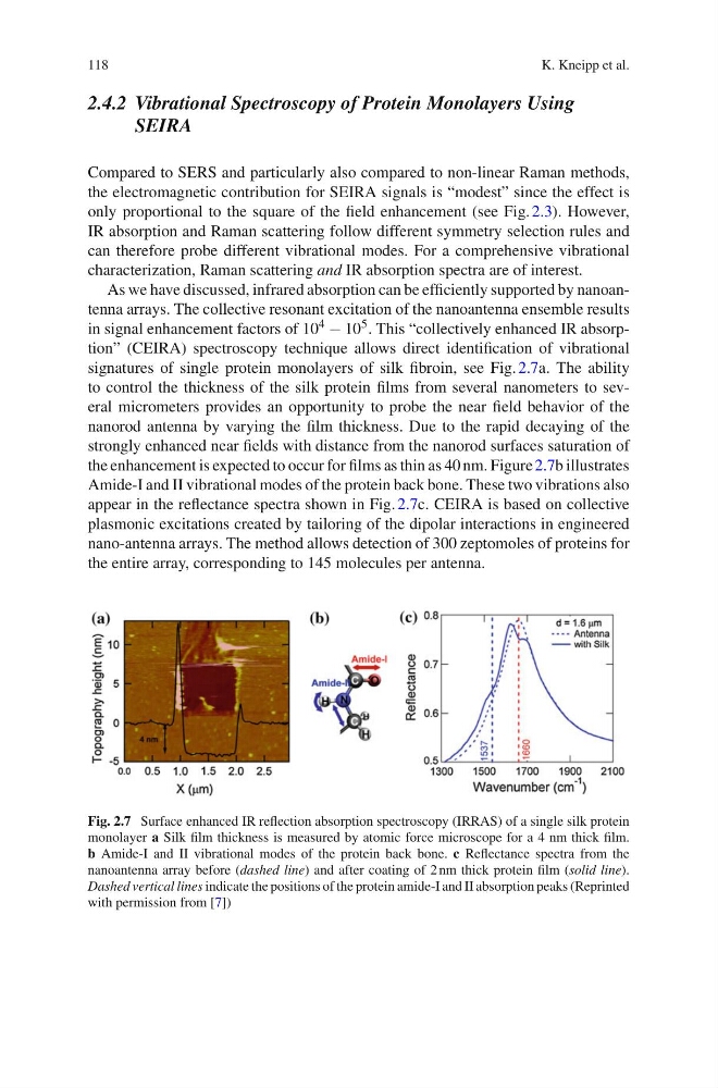
signatures of single protein monolayers of silk fibroin, see Fig. 2.7a. The ability
to control the thickness of the silk protein films from several nanometers to several micrometers provides an opportunity to probe the near field behavior of the
nanorod antenna by varying the film thickness. Due to the rapid decaying of the
strongly enhanced near fields with distance from the nanorod surfaces saturation of
the enhancement is expected to occur for films as thin as 40 nm. Figure 2.7b illustrates
Amide-I and II vibrational modes of the protein back bone. These two vibrations also
appear in the reflectance spectra shown in Fig. 2.7c. CEIRA is based on collective
plasmonic excitations created by tailoring of the dipolar interactions in engineered
nano-antenna arrays. The method allows detection of 300 zeptomoles of proteins for
the entire array, corresponding to 145 molecules per antenna.
Fig. 2.7 Surface enhanced IR reflection absorption spectroscopy (IRRAS) of a single silk protein
monolayer a Silk film thickness is measured by atomic force microscope for a 4 nm thick film.
b Amide-I and II vibrational modes of the protein back bone. c Reflectance spectra from the
nanoantenna array before (dashed line) and after coating of 2 nm thick protein film (solid line).
Dashed vertical lines indicate the positions of the protein amide-I and II absorption peaks (Reprinted
with permission from [7])
K. Kneipp et al.
2.4.2 Vibrational Spectroscopy of Protein Monolayers Using
SEIRA
Compared to SERS and particularly also compared to non-linear Raman methods,
the electromagnetic contribution for SEIRA signals is “modest” since the effect is
only proportional to the square of the field enhancement (see Fig. 2.3). However,
IR absorption and Raman scattering follow different symmetry selection rules and
can therefore probe different vibrational modes. For a comprehensive vibrational
characterization, Raman scattering and IR absorption spectra are of interest.
As we have discussed, infrared absorption can be efficiently supported by nanoantenna arrays. The collective resonant excitation of the nanoantenna ensemble results
in signal enhancement factors of 10 4 − 10 5 . This “collectively enhanced IR absorption” (CEIRA) spectroscopy technique allows direct identification of vibrational
signatures of single protein monolayers of silk fibroin, see Fig. 2.7a. The ability
to control the thickness of the silk protein films from several nanometers to several micrometers provides an opportunity to probe the near field behavior of the
nanorod antenna by varying the film thickness. Due to the rapid decaying of the
strongly enhanced near fields with distance from the nanorod surfaces saturation of
the enhancement is expected to occur for films as thin as 40 nm. Figure 2.7b illustrates
Amide-I and II vibrational modes of the protein back bone. These two vibrations also
appear in the reflectance spectra shown in Fig. 2.7c. CEIRA is based on collective
plasmonic excitations created by tailoring of the dipolar interactions in engineered
nano-antenna arrays. The method allows detection of 300 zeptomoles of proteins for
the entire array, corresponding to 145 molecules per antenna.
Fig. 2.7 Surface enhanced IR reflection absorption spectroscopy (IRRAS) of a single silk protein
monolayer a Silk film thickness is measured by atomic force microscope for a 4 nm thick film.
b Amide-I and II vibrational modes of the protein back bone. c Reflectance spectra from the
nanoantenna array before (dashed line) and after coating of 2 nm thick protein film (solid line).
Dashed vertical lines indicate the positions of the protein amide-I and II absorption peaks (Reprinted
with permission from [7])