

22 NIR Imaging
545
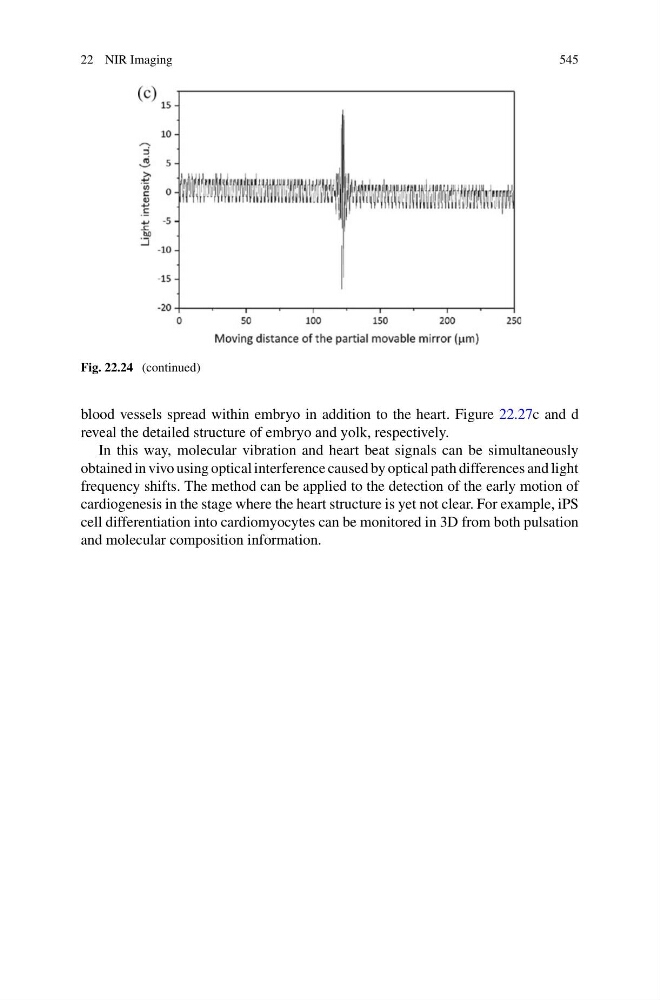
Fig. 22.24 (continued)
blood vessels spread within embryo in addition to the heart. Figure 22.27c and d
reveal the detailed structure of embryo and yolk, respectively.
In this way, molecular vibration and heart beat signals can be simultaneously
obtained in vivo using optical interference caused by optical path differences and light
frequency shifts. The method can be applied to the detection of the early motion of
cardiogenesis in the stage where the heart structure is yet not clear. For example, iPS
cell differentiation into cardiomyocytes can be monitored in 3D from both pulsation
and molecular composition information.
545
Fig. 22.24 (continued)
blood vessels spread within embryo in addition to the heart. Figure 22.27c and d
reveal the detailed structure of embryo and yolk, respectively.
In this way, molecular vibration and heart beat signals can be simultaneously
obtained in vivo using optical interference caused by optical path differences and light
frequency shifts. The method can be applied to the detection of the early motion of
cardiogenesis in the stage where the heart structure is yet not clear. For example, iPS
cell differentiation into cardiomyocytes can be monitored in 3D from both pulsation
and molecular composition information.