

542
D. Ishikawa et al.
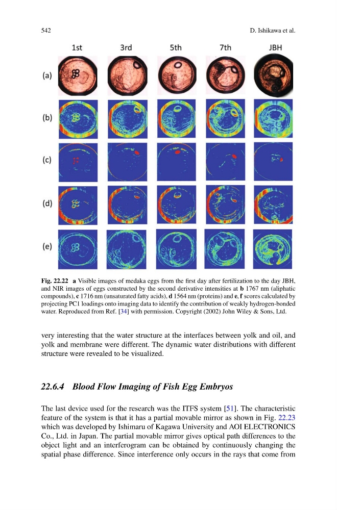
Fig. 22.22 a Visible images of medaka eggs from the first day after fertilization to the day JBH,
and NIR images of eggs constructed by the second derivative intensities at b 1767 nm (aliphatic
compounds), c 1716 nm (unsaturated fatty acids), d 1564 nm (proteins) and e, f scores calculated by
projecting PC1 loadings onto imaging data to identify the contribution of weakly hydrogen-bonded
water. Reproduced from Ref. [34] with permission. Copyright (2002) John Wiley & Sons, Ltd.
very interesting that the water structure at the interfaces between yolk and oil, and
yolk and membrane were different. The dynamic water distributions with different
structure were revealed to be visualized.
22.6.4 Blood Flow Imaging of Fish Egg Embryos
The last device used for the research was the ITFS system [51]. The characteristic
feature of the system is that it has a partial movable mirror as shown in Fig. 22.23
which was developed by Ishimaru of Kagawa University and AOI ELECTRONICS
Co., Ltd. in Japan. The partial movable mirror gives optical path differences to the
object light and an interferogram can be obtained by continuously changing the
spatial phase difference. Since interference only occurs in the rays that come from
D. Ishikawa et al.
Fig. 22.22 a Visible images of medaka eggs from the first day after fertilization to the day JBH,
and NIR images of eggs constructed by the second derivative intensities at b 1767 nm (aliphatic
compounds), c 1716 nm (unsaturated fatty acids), d 1564 nm (proteins) and e, f scores calculated by
projecting PC1 loadings onto imaging data to identify the contribution of weakly hydrogen-bonded
water. Reproduced from Ref. [34] with permission. Copyright (2002) John Wiley & Sons, Ltd.
very interesting that the water structure at the interfaces between yolk and oil, and
yolk and membrane were different. The dynamic water distributions with different
structure were revealed to be visualized.
22.6.4 Blood Flow Imaging of Fish Egg Embryos
The last device used for the research was the ITFS system [51]. The characteristic
feature of the system is that it has a partial movable mirror as shown in Fig. 22.23
which was developed by Ishimaru of Kagawa University and AOI ELECTRONICS
Co., Ltd. in Japan. The partial movable mirror gives optical path differences to the
object light and an interferogram can be obtained by continuously changing the
spatial phase difference. Since interference only occurs in the rays that come from