

20 Medical Applications of NIR Spectroscopy
453
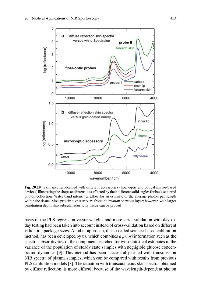
Fig. 20.10 Skin spectra obtained with different accessories (fiber-optic and optical mirror-based
devices) illustrating the shape and intensities affected by their different solid angles for backscattered
photon collection. Water band intensities allow for an estimate of the average photon pathlength
within the tissue. Most protein signatures are from the stratum corneum layer; however, with larger
penetration depth also subcutaneous fatty tissue can be probed
basis of the PLS regression vector weights and more strict validation with day-today testing had been taken into account instead of cross-validation based on different
validation package sizes. Another approach, the so-called science-based calibration
method, has been developed by us, which combines a priori information such as the
spectral absorptivities of the component searched for with statistical estimates of the
variance of the population of steady state samples with negligible glucose concentration dynamics [9]. This method has been successfully tested with transmission
NIR spectra of plasma samples, which can be compared with results from previous
PLS calibration models [8]. The situation with transcutaneous skin spectra, obtained
by diffuse reflection, is more difficult because of the wavelength-dependent photon
453
Fig. 20.10 Skin spectra obtained with different accessories (fiber-optic and optical mirror-based
devices) illustrating the shape and intensities affected by their different solid angles for backscattered
photon collection. Water band intensities allow for an estimate of the average photon pathlength
within the tissue. Most protein signatures are from the stratum corneum layer; however, with larger
penetration depth also subcutaneous fatty tissue can be probed
basis of the PLS regression vector weights and more strict validation with day-today testing had been taken into account instead of cross-validation based on different
validation package sizes. Another approach, the so-called science-based calibration
method, has been developed by us, which combines a priori information such as the
spectral absorptivities of the component searched for with statistical estimates of the
variance of the population of steady state samples with negligible glucose concentration dynamics [9]. This method has been successfully tested with transmission
NIR spectra of plasma samples, which can be compared with results from previous
PLS calibration models [8]. The situation with transcutaneous skin spectra, obtained
by diffuse reflection, is more difficult because of the wavelength-dependent photon