

420
C. W. Huck
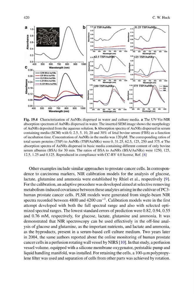
Fig. 19.4 Characterization of AuNRs dispersed in water and culture media. a The UV-Vis-NIR
absorption spectrum of AuNRs dispersed in water. The inserted SEM image shows the morphology
of AuNRs deposited from the aqueous solution. b Absorption spectra of AuNRs dispersed in serum
containing media (SCM) with 0, 2.5, 5, 10, 20 and 30% of fetal bovine serum (FBS) as a function
of incubation time. Concentration of AuNRs in the media was 120 pM. The corresponding ratios of
total serum proteins (TSP) to AuNRs (TSP/AuNRs) were 0, 31.25, 62.5, 125, 250 and 375. c The
absorption spectra of AuNRs dispersed in basic media containing different content of only bovine
serum albumin (BSA) for 30 min. The ratios of BSA to AuNRs (BSA/AuNRs) were 1250, 125,
12.5, 1.25 and 0.125. Reproduced in compliance with CC-BY 4.0 license, Ref. [8]
Other examples include similar approaches to prostate cancer cells. In correspondence to carcinoma markers, NIR calibration models for the analysis of glucose,
lactate, glutamine and ammonia were established by Rhiel et al., respectively [9].
For the calibration, an adaptive procedure was developed aimed at selective removing
metabolism-induced covariance between these analytes arising in the cultivar of PC3human prostate cancer cells. PLSR models were generated from single-beam NIR
spectra recorded between 4800 and 4200 cm
−1 . Calibration models were in the first
attempt developed with both the full spectral range and also with selected optimized spectral ranges. The lowest standard errors of prediction were 0.82, 0.94, 0.55
and 0.76 mM, respectively, for glucose, lactate, glutamine and ammonia. It was
demonstrated that NIR spectroscopy can be used effectively in the off-line analysis of glucose and glutamine, as the important nutrients, and lactate and ammonia,
as the byproducts, present in a serum-based cell culture medium. Two years later
in 2004, the same authors reported about the online monitoring of human prostate
cancer cells in a perfusion rotating wall vessel by NIRS [10]. In that study, a perfusion
vessel volume, equipped with a silicone membrane oxygenator, peristaltic pump and
liquid handling manifold, was installed. For retaining the cells, a 100-μm polypropylene filter was used and separation of cells from other parts was achieved by rotation.
C. W. Huck
Fig. 19.4 Characterization of AuNRs dispersed in water and culture media. a The UV-Vis-NIR
absorption spectrum of AuNRs dispersed in water. The inserted SEM image shows the morphology
of AuNRs deposited from the aqueous solution. b Absorption spectra of AuNRs dispersed in serum
containing media (SCM) with 0, 2.5, 5, 10, 20 and 30% of fetal bovine serum (FBS) as a function
of incubation time. Concentration of AuNRs in the media was 120 pM. The corresponding ratios of
total serum proteins (TSP) to AuNRs (TSP/AuNRs) were 0, 31.25, 62.5, 125, 250 and 375. c The
absorption spectra of AuNRs dispersed in basic media containing different content of only bovine
serum albumin (BSA) for 30 min. The ratios of BSA to AuNRs (BSA/AuNRs) were 1250, 125,
12.5, 1.25 and 0.125. Reproduced in compliance with CC-BY 4.0 license, Ref. [8]
Other examples include similar approaches to prostate cancer cells. In correspondence to carcinoma markers, NIR calibration models for the analysis of glucose,
lactate, glutamine and ammonia were established by Rhiel et al., respectively [9].
For the calibration, an adaptive procedure was developed aimed at selective removing
metabolism-induced covariance between these analytes arising in the cultivar of PC3human prostate cancer cells. PLSR models were generated from single-beam NIR
spectra recorded between 4800 and 4200 cm
−1 . Calibration models were in the first
attempt developed with both the full spectral range and also with selected optimized spectral ranges. The lowest standard errors of prediction were 0.82, 0.94, 0.55
and 0.76 mM, respectively, for glucose, lactate, glutamine and ammonia. It was
demonstrated that NIR spectroscopy can be used effectively in the off-line analysis of glucose and glutamine, as the important nutrients, and lactate and ammonia,
as the byproducts, present in a serum-based cell culture medium. Two years later
in 2004, the same authors reported about the online monitoring of human prostate
cancer cells in a perfusion rotating wall vessel by NIRS [10]. In that study, a perfusion
vessel volume, equipped with a silicone membrane oxygenator, peristaltic pump and
liquid handling manifold, was installed. For retaining the cells, a 100-μm polypropylene filter was used and separation of cells from other parts was achieved by rotation.