

2.1 Holographic Microscopy for the Study of Phase, Diffusive …
65
microobject is recorded, and 3D image reconstructed by it (an optical copy of a
microobject) is studied with the help of a microscope with a resolution of 1 μm
and by a large field of view, by sequential overfocusing of the objective. Sequence
of holograms recorded during the certain period of time allows receiving spatial as
well as time information about the object and thereby characterizing its structurefunctional state. As a rule, during the “lensless” recording of a microobject at the
level of its volumetric image reconstruction, the wave front [91, 138] is used, which
is conjugated with a reference wave front. Then, in the space in front of a hologram
from the side of an observer, the real image is formed, which can be fully studied
on the whole depth of an object at consecutive overfocusing of a microobjective in
spite of its quite short focal distance. Such a method can be used for the studies of
thick layers and separate microobjects which are located in this layer at a great depth.
Virtual images of microobjects reconstructed by a hologram are rarely used. It is due
to the absence of long-focal strong microobjectives and thin substrates, which could
be coated by holographic recording media. Thus, the deeper the object is, the more
difficult it is to study its virtual images.
Another approach of magnification in holographic microscopy is the recording
on a hologram of a microobject an image preliminary enlarged by the adjusted
optical system and located between an object and a hologram [89]. Thus, in this
case, holographing is made not of the objects itself, but of its image. The use of
microobjectives as project optics, which convert the cone of scattered by the object
rays with high numerical aperture into a cone of incident on a hologram rays with
small numerical aperture, allows reducing in comparison with the first approach the
requirements to the photolayer resolution, its density and its homogeneity, but it can
be reached by considerable narrowing of the field of view.
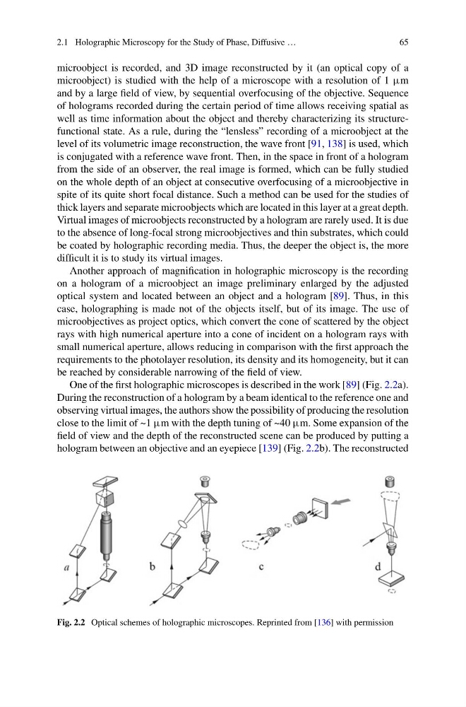
One of the first holographic microscopes is described in the work [89] (Fig. 2.2a).
During the reconstruction of a hologram by a beam identical to the reference one and
observing virtual images, the authors show the possibility of producing the resolution
close to the limit of ~1 μm with the depth tuning of ~40 μm. Some expansion of the
field of view and the depth of the reconstructed scene can be produced by putting a
hologram between an objective and an eyepiece [139] (Fig. 2.2b). The reconstructed
Fig. 2.2 Optical schemes of holographic microscopes. Reprinted from [136] with permission
65
microobject is recorded, and 3D image reconstructed by it (an optical copy of a
microobject) is studied with the help of a microscope with a resolution of 1 μm
and by a large field of view, by sequential overfocusing of the objective. Sequence
of holograms recorded during the certain period of time allows receiving spatial as
well as time information about the object and thereby characterizing its structurefunctional state. As a rule, during the “lensless” recording of a microobject at the
level of its volumetric image reconstruction, the wave front [91, 138] is used, which
is conjugated with a reference wave front. Then, in the space in front of a hologram
from the side of an observer, the real image is formed, which can be fully studied
on the whole depth of an object at consecutive overfocusing of a microobjective in
spite of its quite short focal distance. Such a method can be used for the studies of
thick layers and separate microobjects which are located in this layer at a great depth.
Virtual images of microobjects reconstructed by a hologram are rarely used. It is due
to the absence of long-focal strong microobjectives and thin substrates, which could
be coated by holographic recording media. Thus, the deeper the object is, the more
difficult it is to study its virtual images.
Another approach of magnification in holographic microscopy is the recording
on a hologram of a microobject an image preliminary enlarged by the adjusted
optical system and located between an object and a hologram [89]. Thus, in this
case, holographing is made not of the objects itself, but of its image. The use of
microobjectives as project optics, which convert the cone of scattered by the object
rays with high numerical aperture into a cone of incident on a hologram rays with
small numerical aperture, allows reducing in comparison with the first approach the
requirements to the photolayer resolution, its density and its homogeneity, but it can
be reached by considerable narrowing of the field of view.
One of the first holographic microscopes is described in the work [89] (Fig. 2.2a).
During the reconstruction of a hologram by a beam identical to the reference one and
observing virtual images, the authors show the possibility of producing the resolution
close to the limit of ~1 μm with the depth tuning of ~40 μm. Some expansion of the
field of view and the depth of the reconstructed scene can be produced by putting a
hologram between an objective and an eyepiece [139] (Fig. 2.2b). The reconstructed
Fig. 2.2 Optical schemes of holographic microscopes. Reprinted from [136] with permission