

5.10 Study of Biomechanical Parameters of Skeletal Muscles in …
439
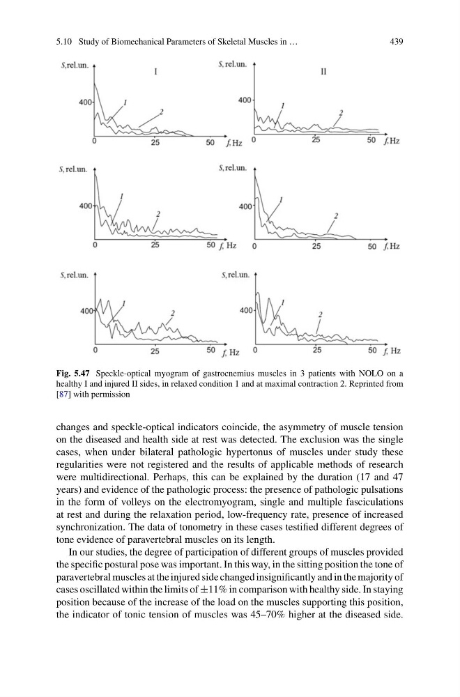
Fig. 5.47 Speckle-optical myogram of gastrocnemius muscles in 3 patients with NOLO on a
healthy I and injured II sides, in relaxed condition 1 and at maximal contraction 2. Reprinted from
[87] with permission
changes and speckle-optical indicators coincide, the asymmetry of muscle tension
on the diseased and health side at rest was detected. The exclusion was the single
cases, when under bilateral pathologic hypertonus of muscles under study these
regularities were not registered and the results of applicable methods of research
were multidirectional. Perhaps, this can be explained by the duration (17 and 47
years) and evidence of the pathologic process: the presence of pathologic pulsations
in the form of volleys on the electromyogram, single and multiple fasciculations
at rest and during the relaxation period, low-frequency rate, presence of increased
synchronization. The data of tonometry in these cases testified different degrees of
tone evidence of paravertebral muscles on its length.
In our studies, the degree of participation of different groups of muscles provided
the specific postural pose was important. In this way, in the sitting position the tone of
paravertebral muscles at the injured side changed insignificantly and in the majority of
cases oscillated within the limits of ±11% in comparison with healthy side. In staying
position because of the increase of the load on the muscles supporting this position,
the indicator of tonic tension of muscles was 45–70% higher at the diseased side.
439
Fig. 5.47 Speckle-optical myogram of gastrocnemius muscles in 3 patients with NOLO on a
healthy I and injured II sides, in relaxed condition 1 and at maximal contraction 2. Reprinted from
[87] with permission
changes and speckle-optical indicators coincide, the asymmetry of muscle tension
on the diseased and health side at rest was detected. The exclusion was the single
cases, when under bilateral pathologic hypertonus of muscles under study these
regularities were not registered and the results of applicable methods of research
were multidirectional. Perhaps, this can be explained by the duration (17 and 47
years) and evidence of the pathologic process: the presence of pathologic pulsations
in the form of volleys on the electromyogram, single and multiple fasciculations
at rest and during the relaxation period, low-frequency rate, presence of increased
synchronization. The data of tonometry in these cases testified different degrees of
tone evidence of paravertebral muscles on its length.
In our studies, the degree of participation of different groups of muscles provided
the specific postural pose was important. In this way, in the sitting position the tone of
paravertebral muscles at the injured side changed insignificantly and in the majority of
cases oscillated within the limits of ±11% in comparison with healthy side. In staying
position because of the increase of the load on the muscles supporting this position,
the indicator of tonic tension of muscles was 45–70% higher at the diseased side.