

212
3 Holographic Interferometry for Studying …
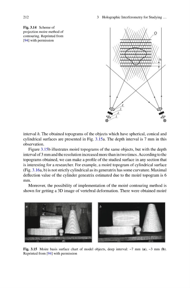
Fig. 3.14 Scheme of
projection moire method of
contouring. Reprinted from
[94] with permission
interval h. The obtained topograms of the objects which have spherical, conical and
cylindrical surfaces are presented in Fig. 3.15a. The depth interval is 7 mm in this
observation.
Figure 3.15b illustrates moiré topograms of the same objects, but with the depth
interval of 3 mm and the resolution increased more than in two times. According to the
topograms obtained, we can make a profile of the studied surface in any section that
is interesting for a researcher. For example, a moiré topogram of cylindrical surface
(Fig. 3.16a, b) is not strictly cylindrical as its generatrix has some curvature. Maximal
deflection value of the cylinder geneatrix estimated due to the moiré topogram is 6
mm.
Moreover, the possibility of implementation of the moiré contouring method is
shown for getting a 3D image of vertebral deformation. There were obtained moiré
Fig. 3.15 Moire basis surface chart of model objects, deep interval: ~7 mm (a), ~3 mm (b).
Reprinted from [94] with permission
3 Holographic Interferometry for Studying …
Fig. 3.14 Scheme of
projection moire method of
contouring. Reprinted from
[94] with permission
interval h. The obtained topograms of the objects which have spherical, conical and
cylindrical surfaces are presented in Fig. 3.15a. The depth interval is 7 mm in this
observation.
Figure 3.15b illustrates moiré topograms of the same objects, but with the depth
interval of 3 mm and the resolution increased more than in two times. According to the
topograms obtained, we can make a profile of the studied surface in any section that
is interesting for a researcher. For example, a moiré topogram of cylindrical surface
(Fig. 3.16a, b) is not strictly cylindrical as its generatrix has some curvature. Maximal
deflection value of the cylinder geneatrix estimated due to the moiré topogram is 6
mm.
Moreover, the possibility of implementation of the moiré contouring method is
shown for getting a 3D image of vertebral deformation. There were obtained moiré
Fig. 3.15 Moire basis surface chart of model objects, deep interval: ~7 mm (a), ~3 mm (b).
Reprinted from [94] with permission