

2.3 Holographic Study with Electrophysiological Control …
145
2.3.4 Study of Singe Isolated Muscle Fibers Under Gaseous
Hyperbary
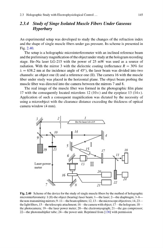
An experimental setup was developed to study the changes of the refraction index
and the shape of single muscle fibers under gas pressure. Its scheme is presented in
Fig. 2.40.
The setup is a holographic microinterferometer with an inclined reference beam
and the preliminary magnification of the object under study at the hologram recording
stage. He–Na laser LG-215 with the power of 25 mW was used as a source of
radiation. With the mirror 3 with the dielectric coating (reflectance R = 30% for
λ = 638.2 nm at the incidence angle of 45°), the laser beam was divided into two
channels: an object one (I) and a reference one (II). The camera 16 with the muscle
fiber under study was placed in the horizontal plane. The object beam probing the
muscle fiber was directed into the camera between the mirrors 7 and 8.
The real image of the muscle fiber was formed in the photographic film plane
17 with the consequently located microlens 12 (10×) and the eyepiece 13 (14×).
Application of such a consequent magnification was dictated by the necessity of
using a microobject with the clearance distance exceeding the thickness of optical
camera window (4 mm).
Fig. 2.40 Scheme of the device for the study of single muscle fibers by the method of holographic
microinterferometry: I (II)-the object (bearing) laser beam; 1—the laser; 2—the diaphragm; 3–8—
the non-transmitting mirrors; 9–11—the beam splitters; 12, 13—the microscope objectives; 14, 23—
the light filters, 15—the telescopic attachment; 16—the camera with object; 17—the hologram; 18—
the photocamera; 19—the laser power meter; 20—the electromyograph; 21—the gas compressor;
22—the photomultiplier tube; 24—the power unit. Reprinted from [136] with permission
145
2.3.4 Study of Singe Isolated Muscle Fibers Under Gaseous
Hyperbary
An experimental setup was developed to study the changes of the refraction index
and the shape of single muscle fibers under gas pressure. Its scheme is presented in
Fig. 2.40.
The setup is a holographic microinterferometer with an inclined reference beam
and the preliminary magnification of the object under study at the hologram recording
stage. He–Na laser LG-215 with the power of 25 mW was used as a source of
radiation. With the mirror 3 with the dielectric coating (reflectance R = 30% for
λ = 638.2 nm at the incidence angle of 45°), the laser beam was divided into two
channels: an object one (I) and a reference one (II). The camera 16 with the muscle
fiber under study was placed in the horizontal plane. The object beam probing the
muscle fiber was directed into the camera between the mirrors 7 and 8.
The real image of the muscle fiber was formed in the photographic film plane
17 with the consequently located microlens 12 (10×) and the eyepiece 13 (14×).
Application of such a consequent magnification was dictated by the necessity of
using a microobject with the clearance distance exceeding the thickness of optical
camera window (4 mm).
Fig. 2.40 Scheme of the device for the study of single muscle fibers by the method of holographic
microinterferometry: I (II)-the object (bearing) laser beam; 1—the laser; 2—the diaphragm; 3–8—
the non-transmitting mirrors; 9–11—the beam splitters; 12, 13—the microscope objectives; 14, 23—
the light filters, 15—the telescopic attachment; 16—the camera with object; 17—the hologram; 18—
the photocamera; 19—the laser power meter; 20—the electromyograph; 21—the gas compressor;
22—the photomultiplier tube; 24—the power unit. Reprinted from [136] with permission