

2.2 Holographic Study of Structural and Functional Characteristics …
117
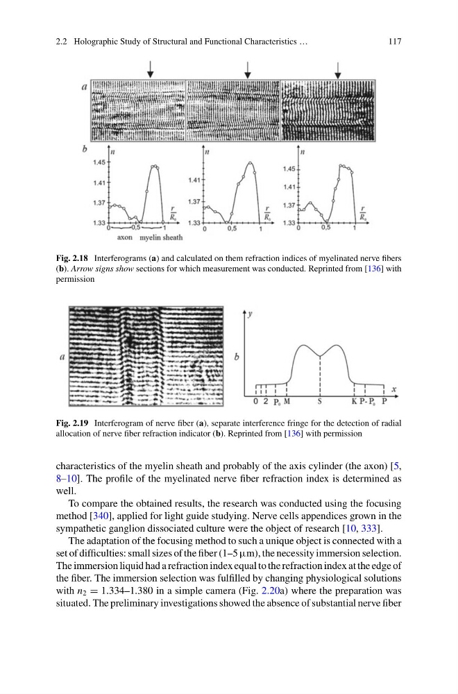
Fig. 2.18 Interferograms (a) and calculated on them refraction indices of myelinated nerve fibers
(b). Arrow signs show sections for which measurement was conducted. Reprinted from [136] with
permission
Fig. 2.19 Interferogram of nerve fiber (a), separate interference fringe for the detection of radial
allocation of nerve fiber refraction indicator (b). Reprinted from [136] with permission
characteristics of the myelin sheath and probably of the axis cylinder (the axon) [5,
8–10]. The profile of the myelinated nerve fiber refraction index is determined as
well.
To compare the obtained results, the research was conducted using the focusing
method [340], applied for light guide studying. Nerve cells appendices grown in the
sympathetic ganglion dissociated culture were the object of research [10, 333].
The adaptation of the focusing method to such a unique object is connected with a
set of difficulties: small sizes of the fiber (1–5 μm), the necessity immersion selection.
The immersion liquid had a refraction index equal to the refraction index at the edge of
the fiber. The immersion selection was fulfilled by changing physiological solutions
with n 2 = 1.334–1.380 in a simple camera (Fig. 2.20a) where the preparation was
situated. The preliminary investigations showed the absence of substantial nerve fiber
117
Fig. 2.18 Interferograms (a) and calculated on them refraction indices of myelinated nerve fibers
(b). Arrow signs show sections for which measurement was conducted. Reprinted from [136] with
permission
Fig. 2.19 Interferogram of nerve fiber (a), separate interference fringe for the detection of radial
allocation of nerve fiber refraction indicator (b). Reprinted from [136] with permission
characteristics of the myelin sheath and probably of the axis cylinder (the axon) [5,
8–10]. The profile of the myelinated nerve fiber refraction index is determined as
well.
To compare the obtained results, the research was conducted using the focusing
method [340], applied for light guide studying. Nerve cells appendices grown in the
sympathetic ganglion dissociated culture were the object of research [10, 333].
The adaptation of the focusing method to such a unique object is connected with a
set of difficulties: small sizes of the fiber (1–5 μm), the necessity immersion selection.
The immersion liquid had a refraction index equal to the refraction index at the edge of
the fiber. The immersion selection was fulfilled by changing physiological solutions
with n 2 = 1.334–1.380 in a simple camera (Fig. 2.20a) where the preparation was
situated. The preliminary investigations showed the absence of substantial nerve fiber