

2.2 Holographic Study of Structural and Functional Characteristics …
79
2.2 Holographic Study of Structural and Functional
Characteristics of Phase Microobjects: Nerve Fibers
and Lymphocytes
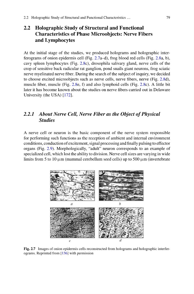
At the initial stage of the studies, we produced holograms and holographic interferograms of onion epidermis cell (Fig. 2.7a–d), frog blood red cells (Fig. 2.8a, b),
cavy spleen lymphocytes (Fig. 2.8c), drosophila salivary gland, nerve cells of the
crop of sensitive back radicular rat ganglion, pond snails giant neurons, frog sciatic
nerve myelinated nerve fiber. During the search of the subject of inquiry, we decided
to choose excited microobjects such as nerve cells, nerve fibers, nerve (Fig. 2.8d),
muscle fiber, muscle (Fig. 2.8e, f) and also lymphoid cells (Fig. 2.8c). A little bit
later it has become known about the studies on nerve fibers carried out in Delaware
University (the USA) [172].
2.2.1 About Nerve Cell, Nerve Fiber as the Object of Physical
Studies
A nerve cell or neuron is the basic component of the nerve system responsible
for performing such functions as the reception of ambient and internal environment
conditions, conduction of excitement, signal processing and finally pulsing to effector
organs (Fig. 2.9). Morphologically, “adult” neuron corresponds to an example of
specialized cell, which lost the ability to division. Nerve cell sizes are varying in wide
limits from 5 to 10 μm (mammal cerebellum seed cells) up to 500 μm (invertebrate
Fig. 2.7 Images of onion epidermis cells reconstructed from holograms and holographic interferograms. Reprinted from [136] with permission
79
2.2 Holographic Study of Structural and Functional
Characteristics of Phase Microobjects: Nerve Fibers
and Lymphocytes
At the initial stage of the studies, we produced holograms and holographic interferograms of onion epidermis cell (Fig. 2.7a–d), frog blood red cells (Fig. 2.8a, b),
cavy spleen lymphocytes (Fig. 2.8c), drosophila salivary gland, nerve cells of the
crop of sensitive back radicular rat ganglion, pond snails giant neurons, frog sciatic
nerve myelinated nerve fiber. During the search of the subject of inquiry, we decided
to choose excited microobjects such as nerve cells, nerve fibers, nerve (Fig. 2.8d),
muscle fiber, muscle (Fig. 2.8e, f) and also lymphoid cells (Fig. 2.8c). A little bit
later it has become known about the studies on nerve fibers carried out in Delaware
University (the USA) [172].
2.2.1 About Nerve Cell, Nerve Fiber as the Object of Physical
Studies
A nerve cell or neuron is the basic component of the nerve system responsible
for performing such functions as the reception of ambient and internal environment
conditions, conduction of excitement, signal processing and finally pulsing to effector
organs (Fig. 2.9). Morphologically, “adult” neuron corresponds to an example of
specialized cell, which lost the ability to division. Nerve cell sizes are varying in wide
limits from 5 to 10 μm (mammal cerebellum seed cells) up to 500 μm (invertebrate
Fig. 2.7 Images of onion epidermis cells reconstructed from holograms and holographic interferograms. Reprinted from [136] with permission