

260
10 Characterization of Atherosclerotic Lesions by Inversion of Eddy-. . .
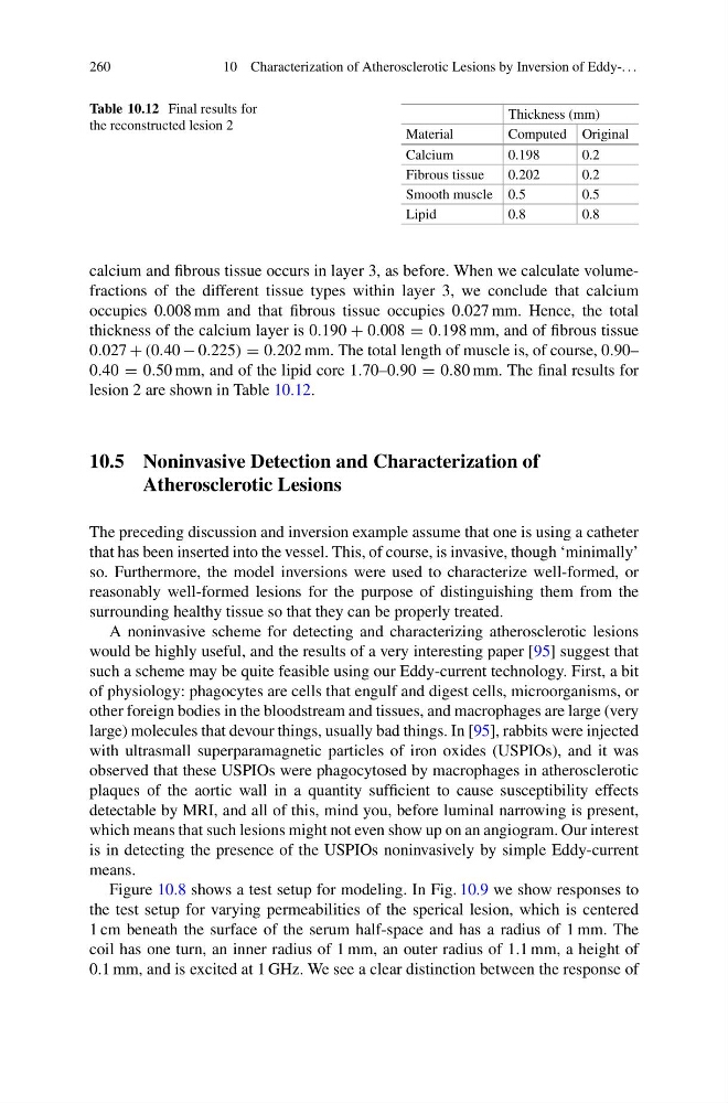
Table 10.12 Final results for
the reconstructed lesion 2
Thickness (mm)
Material
Computed Original
Calcium
0.198
0.2
Fibrous tissue
0.202
0.2
Smooth muscle 0.5
0.5
Lipid
0.8
0.8
calcium and fibrous tissue occurs in layer 3, as before. When we calculate volumefractions of the different tissue types within layer 3, we conclude that calcium
occupies 0.008 mm and that fibrous tissue occupies 0.027 mm. Hence, the total
thickness of the calcium layer is 0.190 + 0.008 = 0.198 mm, and of fibrous tissue
0.027 + (0.40 − 0.225) = 0.202 mm. The total length of muscle is, of course, 0.90–
0.40 = 0.50 mm, and of the lipid core 1.70–0.90 = 0.80 mm. The final results for
lesion 2 are shown in Table 10.12.
10.5 Noninvasive Detection and Characterization of
Atherosclerotic Lesions
The preceding discussion and inversion example assume that one is using a catheter
that has been inserted into the vessel. This, of course, is invasive, though ‘minimally’
so. Furthermore, the model inversions were used to characterize well-formed, or
reasonably well-formed lesions for the purpose of distinguishing them from the
surrounding healthy tissue so that they can be properly treated.
A noninvasive scheme for detecting and characterizing atherosclerotic lesions
would be highly useful, and the results of a very interesting paper [95] suggest that
such a scheme may be quite feasible using our Eddy-current technology. First, a bit
of physiology: phagocytes are cells that engulf and digest cells, microorganisms, or
other foreign bodies in the bloodstream and tissues, and macrophages are large (very
large) molecules that devour things, usually bad things. In [95], rabbits were injected
with ultrasmall superparamagnetic particles of iron oxides (USPIOs), and it was
observed that these USPIOs were phagocytosed by macrophages in atherosclerotic
plaques of the aortic wall in a quantity sufficient to cause susceptibility effects
detectable by MRI, and all of this, mind you, before luminal narrowing is present,
which means that such lesions might not even show up on an angiogram. Our interest
is in detecting the presence of the USPIOs noninvasively by simple Eddy-current
means.
Figure 10.8 shows a test setup for modeling. In Fig. 10.9 we show responses to
the test setup for varying permeabilities of the sperical lesion, which is centered
1 cm beneath the surface of the serum half-space and has a radius of 1 mm. The
coil has one turn, an inner radius of 1 mm, an outer radius of 1.1 mm, a height of
0.1 mm, and is excited at 1 GHz. We see a clear distinction between the response of
10 Characterization of Atherosclerotic Lesions by Inversion of Eddy-. . .
Table 10.12 Final results for
the reconstructed lesion 2
Thickness (mm)
Material
Computed Original
Calcium
0.198
0.2
Fibrous tissue
0.202
0.2
Smooth muscle 0.5
0.5
Lipid
0.8
0.8
calcium and fibrous tissue occurs in layer 3, as before. When we calculate volumefractions of the different tissue types within layer 3, we conclude that calcium
occupies 0.008 mm and that fibrous tissue occupies 0.027 mm. Hence, the total
thickness of the calcium layer is 0.190 + 0.008 = 0.198 mm, and of fibrous tissue
0.027 + (0.40 − 0.225) = 0.202 mm. The total length of muscle is, of course, 0.90–
0.40 = 0.50 mm, and of the lipid core 1.70–0.90 = 0.80 mm. The final results for
lesion 2 are shown in Table 10.12.
10.5 Noninvasive Detection and Characterization of
Atherosclerotic Lesions
The preceding discussion and inversion example assume that one is using a catheter
that has been inserted into the vessel. This, of course, is invasive, though ‘minimally’
so. Furthermore, the model inversions were used to characterize well-formed, or
reasonably well-formed lesions for the purpose of distinguishing them from the
surrounding healthy tissue so that they can be properly treated.
A noninvasive scheme for detecting and characterizing atherosclerotic lesions
would be highly useful, and the results of a very interesting paper [95] suggest that
such a scheme may be quite feasible using our Eddy-current technology. First, a bit
of physiology: phagocytes are cells that engulf and digest cells, microorganisms, or
other foreign bodies in the bloodstream and tissues, and macrophages are large (very
large) molecules that devour things, usually bad things. In [95], rabbits were injected
with ultrasmall superparamagnetic particles of iron oxides (USPIOs), and it was
observed that these USPIOs were phagocytosed by macrophages in atherosclerotic
plaques of the aortic wall in a quantity sufficient to cause susceptibility effects
detectable by MRI, and all of this, mind you, before luminal narrowing is present,
which means that such lesions might not even show up on an angiogram. Our interest
is in detecting the presence of the USPIOs noninvasively by simple Eddy-current
means.
Figure 10.8 shows a test setup for modeling. In Fig. 10.9 we show responses to
the test setup for varying permeabilities of the sperical lesion, which is centered
1 cm beneath the surface of the serum half-space and has a radius of 1 mm. The
coil has one turn, an inner radius of 1 mm, an outer radius of 1.1 mm, a height of
0.1 mm, and is excited at 1 GHz. We see a clear distinction between the response of