

64
M. A. Espinosa-Hernandez et al.
DNA co-modified molecular beacon (MB) and guanine-rich DNA (GDNA) as a
probe. This was made by mixing MB and GDNA at a 1.2:1 ratio inclubated for an
hour at room temperature. Seal probe was prepared by self-templated ligation of 5
-
phosphorylated dumbbell-shaped DNA sequence using T4 DNA ligase. The machine
also required polymerase and nicking enzyme, and a dumbbell-shaped amplification
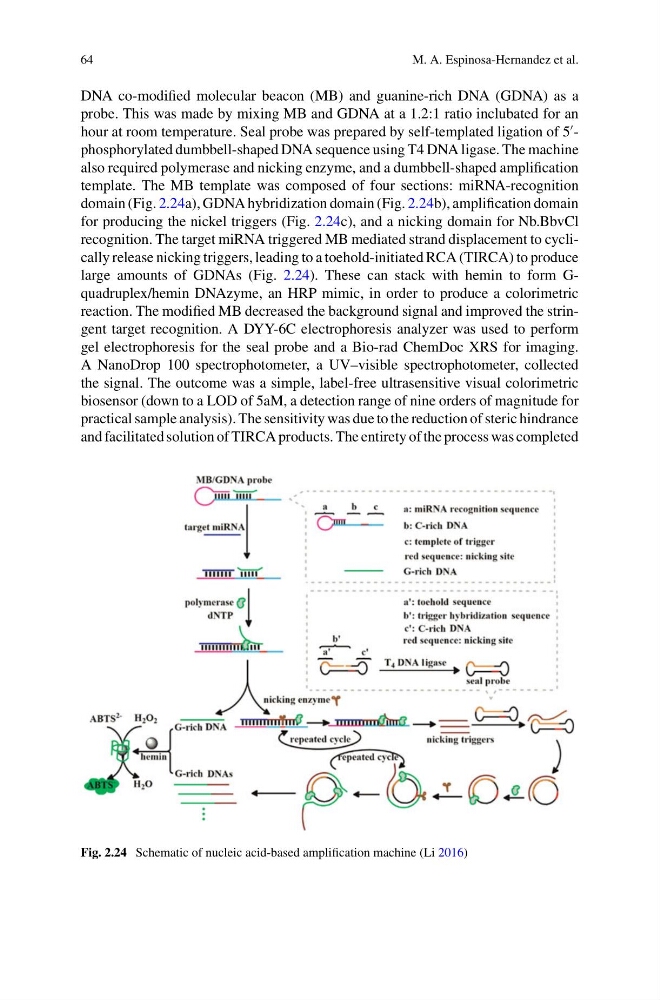
template. The MB template was composed of four sections: miRNA-recognition
domain (Fig. 2.24a), GDNA hybridization domain (Fig. 2.24b), amplification domain
for producing the nickel triggers (Fig. 2.24c), and a nicking domain for Nb.BbvCl
recognition. The target miRNA triggered MB mediated strand displacement to cyclically release nicking triggers, leading to a toehold-initiated RCA (TIRCA) to produce
large amounts of GDNAs (Fig. 2.24). These can stack with hemin to form Gquadruplex/hemin DNAzyme, an HRP mimic, in order to produce a colorimetric
reaction. The modified MB decreased the background signal and improved the stringent target recognition. A DYY-6C electrophoresis analyzer was used to perform
gel electrophoresis for the seal probe and a Bio-rad ChemDoc XRS for imaging.
A NanoDrop 100 spectrophotometer, a UV–visible spectrophotometer, collected
the signal. The outcome was a simple, label-free ultrasensitive visual colorimetric
biosensor (down to a LOD of 5aM, a detection range of nine orders of magnitude for
practical sample analysis). The sensitivity was due to the reduction of steric hindrance
and facilitated solution of TIRCA products. The entirety of the process was completed
Fig. 2.24 Schematic of nucleic acid-based amplification machine (Li 2016)
M. A. Espinosa-Hernandez et al.
DNA co-modified molecular beacon (MB) and guanine-rich DNA (GDNA) as a
probe. This was made by mixing MB and GDNA at a 1.2:1 ratio inclubated for an
hour at room temperature. Seal probe was prepared by self-templated ligation of 5
-
phosphorylated dumbbell-shaped DNA sequence using T4 DNA ligase. The machine
also required polymerase and nicking enzyme, and a dumbbell-shaped amplification
template. The MB template was composed of four sections: miRNA-recognition
domain (Fig. 2.24a), GDNA hybridization domain (Fig. 2.24b), amplification domain
for producing the nickel triggers (Fig. 2.24c), and a nicking domain for Nb.BbvCl
recognition. The target miRNA triggered MB mediated strand displacement to cyclically release nicking triggers, leading to a toehold-initiated RCA (TIRCA) to produce
large amounts of GDNAs (Fig. 2.24). These can stack with hemin to form Gquadruplex/hemin DNAzyme, an HRP mimic, in order to produce a colorimetric
reaction. The modified MB decreased the background signal and improved the stringent target recognition. A DYY-6C electrophoresis analyzer was used to perform
gel electrophoresis for the seal probe and a Bio-rad ChemDoc XRS for imaging.
A NanoDrop 100 spectrophotometer, a UV–visible spectrophotometer, collected
the signal. The outcome was a simple, label-free ultrasensitive visual colorimetric
biosensor (down to a LOD of 5aM, a detection range of nine orders of magnitude for
practical sample analysis). The sensitivity was due to the reduction of steric hindrance
and facilitated solution of TIRCA products. The entirety of the process was completed
Fig. 2.24 Schematic of nucleic acid-based amplification machine (Li 2016)