

40
M. A. Espinosa-Hernandez et al.
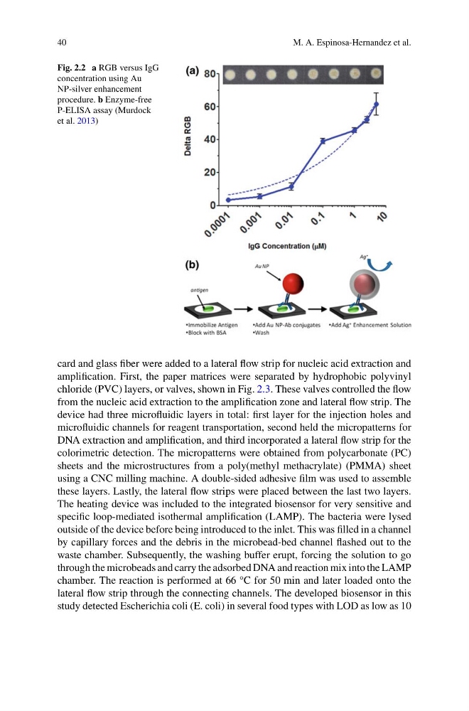
Fig. 2.2 a RGB versus IgG
concentration using Au
NP-silver enhancement
procedure. b Enzyme-free
P-ELISA assay (Murdock
et al. 2013)
card and glass fiber were added to a lateral flow strip for nucleic acid extraction and
amplification. First, the paper matrices were separated by hydrophobic polyvinyl
chloride (PVC) layers, or valves, shown in Fig. 2.3. These valves controlled the flow
from the nucleic acid extraction to the amplification zone and lateral flow strip. The
device had three microfluidic layers in total: first layer for the injection holes and
microfluidic channels for reagent transportation, second held the micropatterns for
DNA extraction and amplification, and third incorporated a lateral flow strip for the
colorimetric detection. The micropatterns were obtained from polycarbonate (PC)
sheets and the microstructures from a poly(methyl methacrylate) (PMMA) sheet
using a CNC milling machine. A double-sided adhesive film was used to assemble
these layers. Lastly, the lateral flow strips were placed between the last two layers.
The heating device was included to the integrated biosensor for very sensitive and
specific loop-mediated isothermal amplification (LAMP). The bacteria were lysed
outside of the device before being introduced to the inlet. This was filled in a channel
by capillary forces and the debris in the microbead-bed channel flashed out to the
waste chamber. Subsequently, the washing buffer erupt, forcing the solution to go
through the microbeads and carry the adsorbed DNA and reaction mix into the LAMP
chamber. The reaction is performed at 66 °C for 50 min and later loaded onto the
lateral flow strip through the connecting channels. The developed biosensor in this
study detected Escherichia coli (E. coli) in several food types with LOD as low as 10
M. A. Espinosa-Hernandez et al.
Fig. 2.2 a RGB versus IgG
concentration using Au
NP-silver enhancement
procedure. b Enzyme-free
P-ELISA assay (Murdock
et al. 2013)
card and glass fiber were added to a lateral flow strip for nucleic acid extraction and
amplification. First, the paper matrices were separated by hydrophobic polyvinyl
chloride (PVC) layers, or valves, shown in Fig. 2.3. These valves controlled the flow
from the nucleic acid extraction to the amplification zone and lateral flow strip. The
device had three microfluidic layers in total: first layer for the injection holes and
microfluidic channels for reagent transportation, second held the micropatterns for
DNA extraction and amplification, and third incorporated a lateral flow strip for the
colorimetric detection. The micropatterns were obtained from polycarbonate (PC)
sheets and the microstructures from a poly(methyl methacrylate) (PMMA) sheet
using a CNC milling machine. A double-sided adhesive film was used to assemble
these layers. Lastly, the lateral flow strips were placed between the last two layers.
The heating device was included to the integrated biosensor for very sensitive and
specific loop-mediated isothermal amplification (LAMP). The bacteria were lysed
outside of the device before being introduced to the inlet. This was filled in a channel
by capillary forces and the debris in the microbead-bed channel flashed out to the
waste chamber. Subsequently, the washing buffer erupt, forcing the solution to go
through the microbeads and carry the adsorbed DNA and reaction mix into the LAMP
chamber. The reaction is performed at 66 °C for 50 min and later loaded onto the
lateral flow strip through the connecting channels. The developed biosensor in this
study detected Escherichia coli (E. coli) in several food types with LOD as low as 10