

24
M. A. Espinosa-Hernandez et al.
Table 2.1
(continued)
BioMEMS platform
Main components
Fabrication strategy Mechanisms of
operation
Detected analyte
Specifics
References
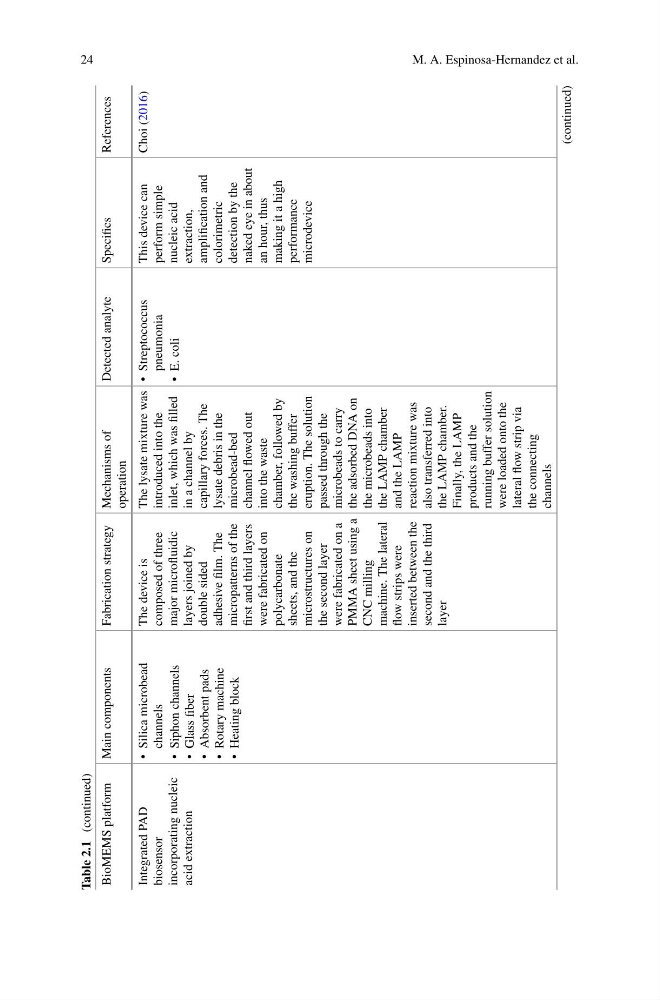
Integrated PAD
biosensor
incorporating nucleic
acid extraction
• Silica microbead
channels
• Siphon channels
• Glass fiber
• Absorbent pads
• Rotary machine
• Heating block
The device is
composed of three
major microfluidic
layers joined by
double sided
adhesive film. The
micropatterns of the
first and third layers
were fabricated on
polycarbonate
sheets, and the
microstructures on
the second layer
were fabricated on a
PMMA sheet using a
CNC milling
machine. The lateral
flow strips were
inserted between the
second and the third
layer
The lysate mixture was
introduced into the
inlet, which was filled
in a channel by
capillary forces. The
lysate debris in the
microbead-bed
channel flowed out
into the waste
chamber, followed by
the washing buffer
eruption. The solution
passed through the
microbeads to carry
the adsorbed DNA on
the microbeads into
the LAMP chamber
and the LAMP
reaction mixture was
also transferred into
the LAMP chamber.
Finally, the LAMP
products and the
running buffer solution
were loaded onto the
lateral flow strip via
the connecting
channels
• Streptococcus
pneumonia
• E. coli
This device can
perform simple
nucleic acid
extraction,
amplification and
colorimetric
detection by the
naked eye in about
an hour, thus
making it a high
performance
microdevice
Choi (2016)
(continued)
M. A. Espinosa-Hernandez et al.
Table 2.1
(continued)
BioMEMS platform
Main components
Fabrication strategy Mechanisms of
operation
Detected analyte
Specifics
References
Integrated PAD
biosensor
incorporating nucleic
acid extraction
• Silica microbead
channels
• Siphon channels
• Glass fiber
• Absorbent pads
• Rotary machine
• Heating block
The device is
composed of three
major microfluidic
layers joined by
double sided
adhesive film. The
micropatterns of the
first and third layers
were fabricated on
polycarbonate
sheets, and the
microstructures on
the second layer
were fabricated on a
PMMA sheet using a
CNC milling
machine. The lateral
flow strips were
inserted between the
second and the third
layer
The lysate mixture was
introduced into the
inlet, which was filled
in a channel by
capillary forces. The
lysate debris in the
microbead-bed
channel flowed out
into the waste
chamber, followed by
the washing buffer
eruption. The solution
passed through the
microbeads to carry
the adsorbed DNA on
the microbeads into
the LAMP chamber
and the LAMP
reaction mixture was
also transferred into
the LAMP chamber.
Finally, the LAMP
products and the
running buffer solution
were loaded onto the
lateral flow strip via
the connecting
channels
• Streptococcus
pneumonia
• E. coli
This device can
perform simple
nucleic acid
extraction,
amplification and
colorimetric
detection by the
naked eye in about
an hour, thus
making it a high
performance
microdevice
Choi (2016)
(continued)