

148
A. S. Cerda-Kipper and S. Hosseini
water-soluble carboxymethylated β-1,3-glucan (CMG) as a long spacer arm (LSA)
coupled with aminated DNA-capture probes on magnetic particles (MPs). Significant enhancements were observed in DNA hybridization thanks to employment of
the LSA-functionalized magnetic particles (LSA-MPs) due to the freedom that their
long and flexible spacer arms provide for DNA hybridization close to the MP surface.
It was observed that a limited sensitivity can be achieved due to the attenuation of
CL by MPs. The authors found that, in order to allow the target to be independently
detected while preventing the attenuation in CL intensity, the CL labels should be
released from MPs. This shortcoming was addressed by adapting the new strategy
of using covalent attachment to link the LSA-containing capture probes to MPs and
resultant hybridization with target DNA fragments altered with CL labels, anticipating the release form the LSA-MPs upon LSA fracture or DNA degradation. The
authors performed two methods for the release of CL labels from the LSA-MPs: DNA
enzymolysis and LSA ultrasonication. The cleaned, magnetic complexes were resuspended PBS buffer and treated with ultrasonic vibration, respectively. Since DNase
I nonspecifically degrades double- or single-stranded DNA to mononucleotides or
oligonucleotides, Deoxyribonuclease (DNase) I was utilized for degradation of DNA
to release the labels. The genomic DNA/RNA of a target pathogen was separated
from the patient’s serum, and through RT-PCR or PCR using biotin-11-dUTP, biotinylated amplicons were generated. Consequently, LSA-MPs were used to capture the
biotinylated amplicons and combined with streptavidin–alkaline phosphatase (SAAP). Treatment was performed with ultrasonic vibration or DNase and magnetic
separation, the resultant CL labels (AP tags) converted the substrate AMPPD to
AMP-D, catalytically and when exposed to the emission of a prolonged, strong CL
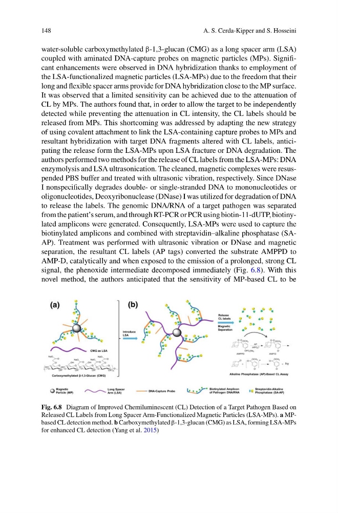
signal, the phenoxide intermediate decomposed immediately (Fig. 6.8). With this
novel method, the authors anticipated that the sensitivity of MP-based CL to be
Fig. 6.8 Diagram of Improved Chemiluminescent (CL) Detection of a Target Pathogen Based on
Released CL Labels from Long Spacer Arm-Functionalized Magnetic Particles (LSA-MPs). a MPbased CL detection method. b Carboxymethylated β-1,3-glucan (CMG) as LSA, forming LSA-MPs
for enhanced CL detection (Yang et al. 2015)
A. S. Cerda-Kipper and S. Hosseini
water-soluble carboxymethylated β-1,3-glucan (CMG) as a long spacer arm (LSA)
coupled with aminated DNA-capture probes on magnetic particles (MPs). Significant enhancements were observed in DNA hybridization thanks to employment of
the LSA-functionalized magnetic particles (LSA-MPs) due to the freedom that their
long and flexible spacer arms provide for DNA hybridization close to the MP surface.
It was observed that a limited sensitivity can be achieved due to the attenuation of
CL by MPs. The authors found that, in order to allow the target to be independently
detected while preventing the attenuation in CL intensity, the CL labels should be
released from MPs. This shortcoming was addressed by adapting the new strategy
of using covalent attachment to link the LSA-containing capture probes to MPs and
resultant hybridization with target DNA fragments altered with CL labels, anticipating the release form the LSA-MPs upon LSA fracture or DNA degradation. The
authors performed two methods for the release of CL labels from the LSA-MPs: DNA
enzymolysis and LSA ultrasonication. The cleaned, magnetic complexes were resuspended PBS buffer and treated with ultrasonic vibration, respectively. Since DNase
I nonspecifically degrades double- or single-stranded DNA to mononucleotides or
oligonucleotides, Deoxyribonuclease (DNase) I was utilized for degradation of DNA
to release the labels. The genomic DNA/RNA of a target pathogen was separated
from the patient’s serum, and through RT-PCR or PCR using biotin-11-dUTP, biotinylated amplicons were generated. Consequently, LSA-MPs were used to capture the
biotinylated amplicons and combined with streptavidin–alkaline phosphatase (SAAP). Treatment was performed with ultrasonic vibration or DNase and magnetic
separation, the resultant CL labels (AP tags) converted the substrate AMPPD to
AMP-D, catalytically and when exposed to the emission of a prolonged, strong CL
signal, the phenoxide intermediate decomposed immediately (Fig. 6.8). With this
novel method, the authors anticipated that the sensitivity of MP-based CL to be
Fig. 6.8 Diagram of Improved Chemiluminescent (CL) Detection of a Target Pathogen Based on
Released CL Labels from Long Spacer Arm-Functionalized Magnetic Particles (LSA-MPs). a MPbased CL detection method. b Carboxymethylated β-1,3-glucan (CMG) as LSA, forming LSA-MPs
for enhanced CL detection (Yang et al. 2015)