

4.2 Eukaryotic Flagella and Cilia
71
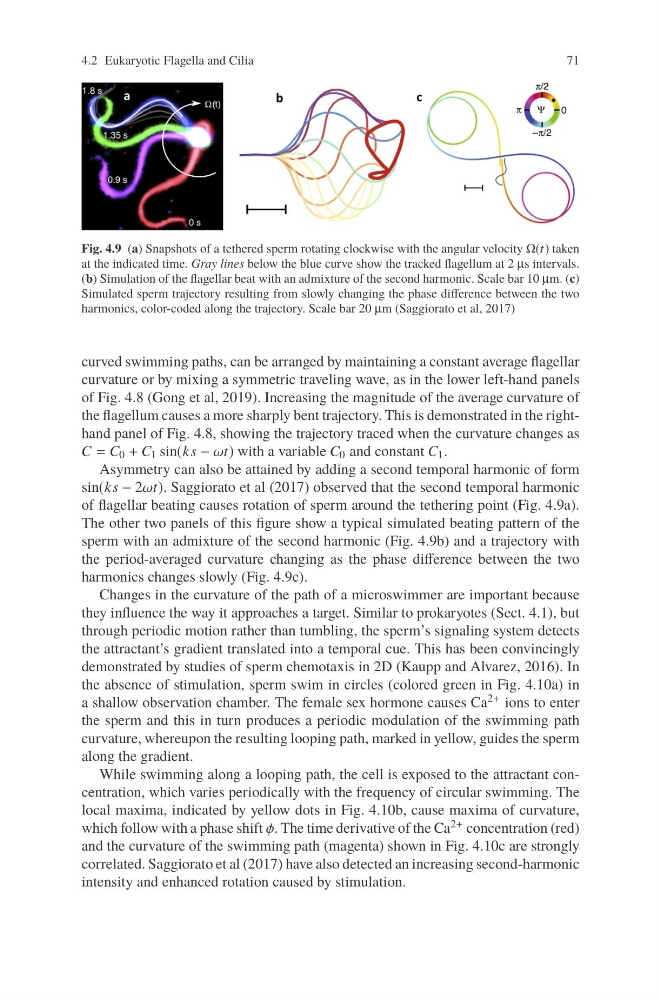
Fig. 4.9 (a) Snapshots of a tethered sperm rotating clockwise with the angular velocity Ω(t) taken
at the indicated time. Gray lines below the blue curve show the tracked flagellum at 2 μs intervals.
(b) Simulation of the flagellar beat with an admixture of the second harmonic. Scale bar 10 μm. (c)
Simulated sperm trajectory resulting from slowly changing the phase difference between the two
harmonics, color-coded along the trajectory. Scale bar 20 μm (Saggiorato et al, 2017)
curved swimming paths, can be arranged by maintaining a constant average flagellar
curvature or by mixing a symmetric traveling wave, as in the lower left-hand panels
of Fig. 4.8 (Gong et al, 2019). Increasing the magnitude of the average curvature of
the flagellum causes a more sharply bent trajectory. This is demonstrated in the righthand panel of Fig. 4.8, showing the trajectory traced when the curvature changes as
C = C 0 + C 1 sin(ks − ωt) with a variable C 0 and constant C 1 .
Asymmetry can also be attained by adding a second temporal harmonic of form
sin(ks − 2ωt). Saggiorato et al (2017) observed that the second temporal harmonic
of flagellar beating causes rotation of sperm around the tethering point (Fig. 4.9a).
The other two panels of this figure show a typical simulated beating pattern of the
sperm with an admixture of the second harmonic (Fig. 4.9b) and a trajectory with
the period-averaged curvature changing as the phase difference between the two
harmonics changes slowly (Fig. 4.9c).
Changes in the curvature of the path of a microswimmer are important because
they influence the way it approaches a target. Similar to prokaryotes (Sect. 4.1), but
through periodic motion rather than tumbling, the sperm’s signaling system detects
the attractant’s gradient translated into a temporal cue. This has been convincingly
demonstrated by studies of sperm chemotaxis in 2D (Kaupp and Alvarez, 2016). In
the absence of stimulation, sperm swim in circles (colored green in Fig. 4.10a) in
a shallow observation chamber. The female sex hormone causes Ca 2+ ions to enter
the sperm and this in turn produces a periodic modulation of the swimming path
curvature, whereupon the resulting looping path, marked in yellow, guides the sperm
along the gradient.
While swimming along a looping path, the cell is exposed to the attractant concentration, which varies periodically with the frequency of circular swimming. The
local maxima, indicated by yellow dots in Fig. 4.10b, cause maxima of curvature,
which follow with a phase shift φ. The time derivative of the Ca 2+ concentration (red)
and the curvature of the swimming path (magenta) shown in Fig. 4.10c are strongly
correlated. Saggiorato et al (2017) have also detected an increasing second-harmonic
intensity and enhanced rotation caused by stimulation.
71
Fig. 4.9 (a) Snapshots of a tethered sperm rotating clockwise with the angular velocity Ω(t) taken
at the indicated time. Gray lines below the blue curve show the tracked flagellum at 2 μs intervals.
(b) Simulation of the flagellar beat with an admixture of the second harmonic. Scale bar 10 μm. (c)
Simulated sperm trajectory resulting from slowly changing the phase difference between the two
harmonics, color-coded along the trajectory. Scale bar 20 μm (Saggiorato et al, 2017)
curved swimming paths, can be arranged by maintaining a constant average flagellar
curvature or by mixing a symmetric traveling wave, as in the lower left-hand panels
of Fig. 4.8 (Gong et al, 2019). Increasing the magnitude of the average curvature of
the flagellum causes a more sharply bent trajectory. This is demonstrated in the righthand panel of Fig. 4.8, showing the trajectory traced when the curvature changes as
C = C 0 + C 1 sin(ks − ωt) with a variable C 0 and constant C 1 .
Asymmetry can also be attained by adding a second temporal harmonic of form
sin(ks − 2ωt). Saggiorato et al (2017) observed that the second temporal harmonic
of flagellar beating causes rotation of sperm around the tethering point (Fig. 4.9a).
The other two panels of this figure show a typical simulated beating pattern of the
sperm with an admixture of the second harmonic (Fig. 4.9b) and a trajectory with
the period-averaged curvature changing as the phase difference between the two
harmonics changes slowly (Fig. 4.9c).
Changes in the curvature of the path of a microswimmer are important because
they influence the way it approaches a target. Similar to prokaryotes (Sect. 4.1), but
through periodic motion rather than tumbling, the sperm’s signaling system detects
the attractant’s gradient translated into a temporal cue. This has been convincingly
demonstrated by studies of sperm chemotaxis in 2D (Kaupp and Alvarez, 2016). In
the absence of stimulation, sperm swim in circles (colored green in Fig. 4.10a) in
a shallow observation chamber. The female sex hormone causes Ca 2+ ions to enter
the sperm and this in turn produces a periodic modulation of the swimming path
curvature, whereupon the resulting looping path, marked in yellow, guides the sperm
along the gradient.
While swimming along a looping path, the cell is exposed to the attractant concentration, which varies periodically with the frequency of circular swimming. The
local maxima, indicated by yellow dots in Fig. 4.10b, cause maxima of curvature,
which follow with a phase shift φ. The time derivative of the Ca 2+ concentration (red)
and the curvature of the swimming path (magenta) shown in Fig. 4.10c are strongly
correlated. Saggiorato et al (2017) have also detected an increasing second-harmonic
intensity and enhanced rotation caused by stimulation.