

4.2 Eukaryotic Flagella and Cilia
69
over, swirling flow due to flagellar rotation, as shown in the right panels of Fig. 4.3,
leads to their wrapping. Nevertheless, the filaments do not get tangled up since all
of them coil in the same direction.
4.2 Eukaryotic Flagella and Cilia
Eukaryotic flagella do not rotate, but they can beat in various fashions; sperm also
propels itself in this way. Their core is a microtubule-based cytoskeletal structure
called an axoneme. More variegated beating patterns are possible because molecular
motors, called axonemal dyneins, are distributed along the axoneme rather than being
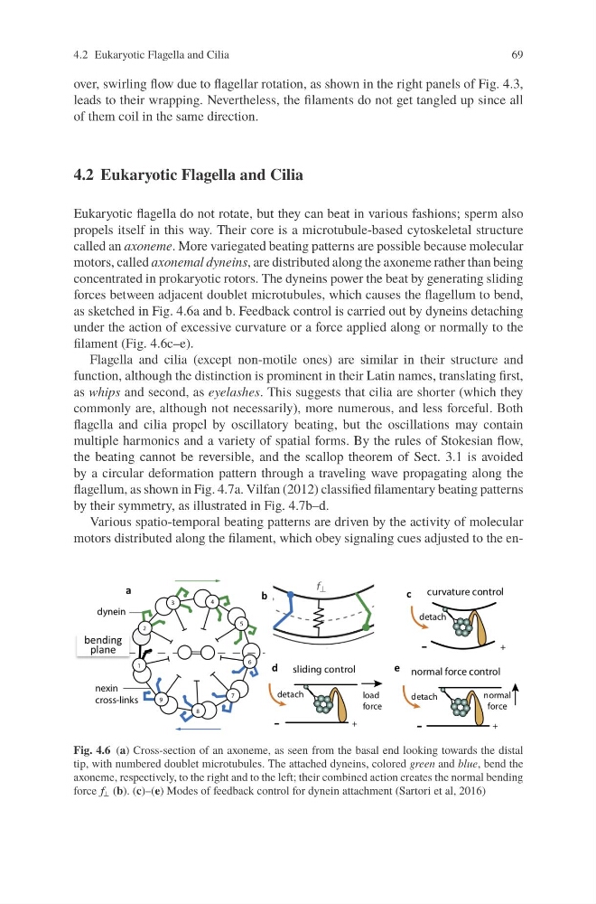
concentrated in prokaryotic rotors. The dyneins power the beat by generating sliding
forces between adjacent doublet microtubules, which causes the flagellum to bend,
as sketched in Fig. 4.6a and b. Feedback control is carried out by dyneins detaching
under the action of excessive curvature or a force applied along or normally to the
filament (Fig. 4.6c–e).
Flagella and cilia (except non-motile ones) are similar in their structure and
function, although the distinction is prominent in their Latin names, translating first,
as whips and second, as eyelashes. This suggests that cilia are shorter (which they
commonly are, although not necessarily), more numerous, and less forceful. Both
flagella and cilia propel by oscillatory beating, but the oscillations may contain
multiple harmonics and a variety of spatial forms. By the rules of Stokesian flow,
the beating cannot be reversible, and the scallop theorem of Sect. 3.1 is avoided
by a circular deformation pattern through a traveling wave propagating along the
flagellum, as shown in Fig. 4.7a. Vilfan (2012) classified filamentary beating patterns
by their symmetry, as illustrated in Fig. 4.7b–d.
Various spatio-temporal beating patterns are driven by the activity of molecular
motors distributed along the filament, which obey signaling cues adjusted to the enFig. 4.6 (a) Cross-section of an axoneme, as seen from the basal end looking towards the distal
tip, with numbered doublet microtubules. The attached dyneins, colored green and blue, bend the
axoneme, respectively, to the right and to the left; their combined action creates the normal bending
force f ⊥ (b). (c)–(e) Modes of feedback control for dynein attachment (Sartori et al, 2016)
69
over, swirling flow due to flagellar rotation, as shown in the right panels of Fig. 4.3,
leads to their wrapping. Nevertheless, the filaments do not get tangled up since all
of them coil in the same direction.
4.2 Eukaryotic Flagella and Cilia
Eukaryotic flagella do not rotate, but they can beat in various fashions; sperm also
propels itself in this way. Their core is a microtubule-based cytoskeletal structure
called an axoneme. More variegated beating patterns are possible because molecular
motors, called axonemal dyneins, are distributed along the axoneme rather than being
concentrated in prokaryotic rotors. The dyneins power the beat by generating sliding
forces between adjacent doublet microtubules, which causes the flagellum to bend,
as sketched in Fig. 4.6a and b. Feedback control is carried out by dyneins detaching
under the action of excessive curvature or a force applied along or normally to the
filament (Fig. 4.6c–e).
Flagella and cilia (except non-motile ones) are similar in their structure and
function, although the distinction is prominent in their Latin names, translating first,
as whips and second, as eyelashes. This suggests that cilia are shorter (which they
commonly are, although not necessarily), more numerous, and less forceful. Both
flagella and cilia propel by oscillatory beating, but the oscillations may contain
multiple harmonics and a variety of spatial forms. By the rules of Stokesian flow,
the beating cannot be reversible, and the scallop theorem of Sect. 3.1 is avoided
by a circular deformation pattern through a traveling wave propagating along the
flagellum, as shown in Fig. 4.7a. Vilfan (2012) classified filamentary beating patterns
by their symmetry, as illustrated in Fig. 4.7b–d.
Various spatio-temporal beating patterns are driven by the activity of molecular
motors distributed along the filament, which obey signaling cues adjusted to the enFig. 4.6 (a) Cross-section of an axoneme, as seen from the basal end looking towards the distal
tip, with numbered doublet microtubules. The attached dyneins, colored green and blue, bend the
axoneme, respectively, to the right and to the left; their combined action creates the normal bending
force f ⊥ (b). (c)–(e) Modes of feedback control for dynein attachment (Sartori et al, 2016)