

66
4 Motion of Microorganisms
by steps in the direction of rotation. The motor is powered electrochemically, using
energy from either sodium or hydrogen ion flux through the membrane – this is
the way things usually work in prokaryotic cells, taken over by eukaryotes in their
mitochondria. Experimentalists have succeeded in following the stepped activation
sequence of protein units, and have been able to reduce the motive force by lowering
the ion concentration, which led to a decrease in both the number of stator units and
the speed per unit (Sowa et al, 2005).
A helical filament is a cylinder formed by 11 protofilaments built of a stack
of flagellin protein monomers, which can assume either left-hand or right-hand
(L-R) conformations differing in length. Each protofilament contains a single type
of monomer, and therefore L- and R-protofilaments have different lengths. Mixing
them in the flagellar filament produces bending which, combined with an intrinsic
twist of the filament, leads to a helical configuration with the pitch decreasing as
the difference between the number of protofilaments with different conformations is
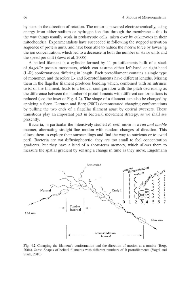
reduced (see the inset of Fig. 4.2). The shape of a filament can also be changed by
applying a force. Darnton and Berg (2007) demonstrated changing conformations
by pulling the two ends of a flagellar filament apart by optical tweezers. These
transitions play an important part in bacterial movement strategy, as we shall see
presently.
Bacteria, in particular the intensively studied E. coli, move in a run and tumble
manner, alternating straight-line motion with random changes of direction. This
allows them to explore their surroundings and find the way to nutrients or to avoid
peril. Bacteria are not diffusiophoretic: they are too small to feel concentration
gradients, but they have a kind of a short-term memory, which allows them to
measure the spatial gradient by sensing a change in time as they move. Engelmann
Fig. 4.2 Changing the filament’s conformation and the direction of motion at a tumble (Berg,
2004). Inset: Shapes of helical filaments with different numbers of R-protofilaments (Vogel and
Stark, 2010)
4 Motion of Microorganisms
by steps in the direction of rotation. The motor is powered electrochemically, using
energy from either sodium or hydrogen ion flux through the membrane – this is
the way things usually work in prokaryotic cells, taken over by eukaryotes in their
mitochondria. Experimentalists have succeeded in following the stepped activation
sequence of protein units, and have been able to reduce the motive force by lowering
the ion concentration, which led to a decrease in both the number of stator units and
the speed per unit (Sowa et al, 2005).
A helical filament is a cylinder formed by 11 protofilaments built of a stack
of flagellin protein monomers, which can assume either left-hand or right-hand
(L-R) conformations differing in length. Each protofilament contains a single type
of monomer, and therefore L- and R-protofilaments have different lengths. Mixing
them in the flagellar filament produces bending which, combined with an intrinsic
twist of the filament, leads to a helical configuration with the pitch decreasing as
the difference between the number of protofilaments with different conformations is
reduced (see the inset of Fig. 4.2). The shape of a filament can also be changed by
applying a force. Darnton and Berg (2007) demonstrated changing conformations
by pulling the two ends of a flagellar filament apart by optical tweezers. These
transitions play an important part in bacterial movement strategy, as we shall see
presently.
Bacteria, in particular the intensively studied E. coli, move in a run and tumble
manner, alternating straight-line motion with random changes of direction. This
allows them to explore their surroundings and find the way to nutrients or to avoid
peril. Bacteria are not diffusiophoretic: they are too small to feel concentration
gradients, but they have a kind of a short-term memory, which allows them to
measure the spatial gradient by sensing a change in time as they move. Engelmann
Fig. 4.2 Changing the filament’s conformation and the direction of motion at a tumble (Berg,
2004). Inset: Shapes of helical filaments with different numbers of R-protofilaments (Vogel and
Stark, 2010)