

8.6 Remodeling
189
surface mechanics and cell sorting in development is reviewed by Lecuit and Lenne
(2007) and Krens and Heisenberg (2011).
The crucial event requiring radical remodeling of the embryonic tissue is gastrulation, turning a plain hollow sheath of cells, blastula, into a layered structure,
gastrula. Lewis Wolpert called it “truly the most important time in your life”. The site
of invagination and internalization of the cells driven to inner layers is designated by
a furrow formed by apical constriction of ventral cells. This is a standard mechanism
of epithelial folding that involves, as noted in Sect. 7.7, enhanced actomyosin activity. However, the development of the ventral furrow in Drosophila does not proceed
continuously, but involves repeated pulsed constrictions, which are asynchronous
between neighboring cells (Martin et al, 2009). As shown in Fig. 8.18, individual
cells go through the stages of constriction and stabilization, while the average apical area of a band of cells oscillates as well. These pulsations are accompanied by
oscillations of myosin intensity, both on the average and in individual cells.
Gastrulation is necessarily accompanied by global tissue deformations. No in
vivo force measurements are available as yet, but Streichan et al (2018) have mapped
cellular flow together with the accompanying myosin intensity, as an indicator of
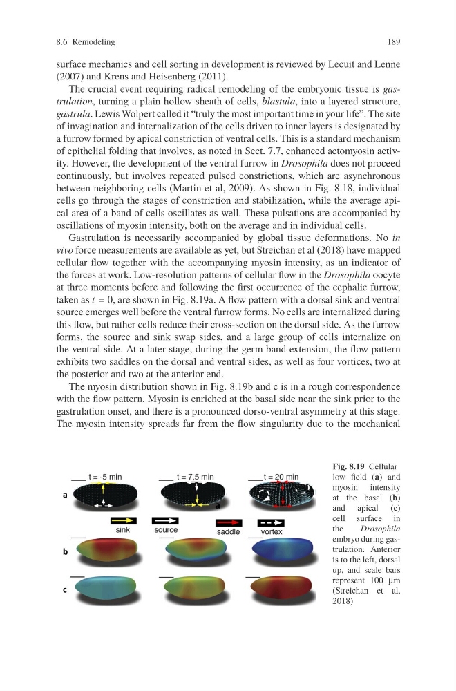
the forces at work. Low-resolution patterns of cellular flow in the Drosophila oocyte
at three moments before and following the first occurrence of the cephalic furrow,
taken as t = 0, are shown in Fig. 8.19a. A flow pattern with a dorsal sink and ventral
source emerges well before the ventral furrow forms. No cells are internalized during
this flow, but rather cells reduce their cross-section on the dorsal side. As the furrow
forms, the source and sink swap sides, and a large group of cells internalize on
the ventral side. At a later stage, during the germ band extension, the flow pattern
exhibits two saddles on the dorsal and ventral sides, as well as four vortices, two at
the posterior and two at the anterior end.
The myosin distribution shown in Fig. 8.19b and c is in a rough correspondence
with the flow pattern. Myosin is enriched at the basal side near the sink prior to the
gastrulation onset, and there is a pronounced dorso-ventral asymmetry at this stage.
The myosin intensity spreads far from the flow singularity due to the mechanical
Fig. 8.19 Cellular
low field (a) and
myosin intensity
at the basal (b)
and apical (c)
cell surface in
the
Drosophila
embryo during gastrulation. Anterior
is to the left, dorsal
up, and scale bars
represent 100 μm
(Streichan et al,
2018)
189
surface mechanics and cell sorting in development is reviewed by Lecuit and Lenne
(2007) and Krens and Heisenberg (2011).
The crucial event requiring radical remodeling of the embryonic tissue is gastrulation, turning a plain hollow sheath of cells, blastula, into a layered structure,
gastrula. Lewis Wolpert called it “truly the most important time in your life”. The site
of invagination and internalization of the cells driven to inner layers is designated by
a furrow formed by apical constriction of ventral cells. This is a standard mechanism
of epithelial folding that involves, as noted in Sect. 7.7, enhanced actomyosin activity. However, the development of the ventral furrow in Drosophila does not proceed
continuously, but involves repeated pulsed constrictions, which are asynchronous
between neighboring cells (Martin et al, 2009). As shown in Fig. 8.18, individual
cells go through the stages of constriction and stabilization, while the average apical area of a band of cells oscillates as well. These pulsations are accompanied by
oscillations of myosin intensity, both on the average and in individual cells.
Gastrulation is necessarily accompanied by global tissue deformations. No in
vivo force measurements are available as yet, but Streichan et al (2018) have mapped
cellular flow together with the accompanying myosin intensity, as an indicator of
the forces at work. Low-resolution patterns of cellular flow in the Drosophila oocyte
at three moments before and following the first occurrence of the cephalic furrow,
taken as t = 0, are shown in Fig. 8.19a. A flow pattern with a dorsal sink and ventral
source emerges well before the ventral furrow forms. No cells are internalized during
this flow, but rather cells reduce their cross-section on the dorsal side. As the furrow
forms, the source and sink swap sides, and a large group of cells internalize on
the ventral side. At a later stage, during the germ band extension, the flow pattern
exhibits two saddles on the dorsal and ventral sides, as well as four vortices, two at
the posterior and two at the anterior end.
The myosin distribution shown in Fig. 8.19b and c is in a rough correspondence
with the flow pattern. Myosin is enriched at the basal side near the sink prior to the
gastrulation onset, and there is a pronounced dorso-ventral asymmetry at this stage.
The myosin intensity spreads far from the flow singularity due to the mechanical
Fig. 8.19 Cellular
low field (a) and
myosin intensity
at the basal (b)
and apical (c)
cell surface in
the
Drosophila
embryo during gastrulation. Anterior
is to the left, dorsal
up, and scale bars
represent 100 μm
(Streichan et al,
2018)